Volume 12, Number 4—April 2006
Dispatch
Avian Influenza H5N1 in Naturally Infected Domestic Cat
Thaweesak Songserm*, Alongkorn Amonsin†, Rungroj Jam-on*, Namdee Sae-Heng*, Noppadol Meemak‡, Nuananong Pariyothorn†, Sunchai Payungporn†, Apiradee Theamboonlers†, and Yong Poovorawan†
Figure 1
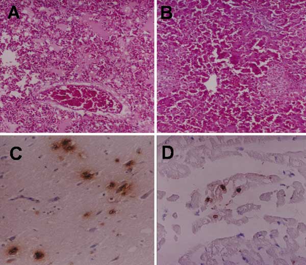
Figure 1. Microscopic lesions of the infected cat, lung edema with homogeneous pink material and congestion (A) and multifocal necrosis in the liver (B). Positive sites are shown by immunohistochemical examination of the infected cat in neurons (C) and cardiac muscle cells (D) (magnification ×100).
Page created: January 23, 2012
Page updated: January 23, 2012
Page reviewed: January 23, 2012
The conclusions, findings, and opinions expressed by authors contributing to this journal do not necessarily reflect the official position of the U.S. Department of Health and Human Services, the Public Health Service, the Centers for Disease Control and Prevention, or the authors' affiliated institutions. Use of trade names is for identification only and does not imply endorsement by any of the groups named above.