Volume 12, Number 7—July 2006
Research
Epidemiologic and Molecular Analysis of Human Tularemia, United States, 1964–2004
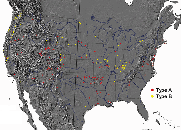
Figure 1

Figure 1. Geographic distribution for type A (red circles) and type B (yellow circles) Francisella tularensis isolates from humans, contiguous United States, 1964–2004. Each circle represents 1 isolate. Isolates were plotted randomly within the county of exposure. The 100th meridian is indicated by the black line transecting the United States. County of exposure was known for 198 (63%) isolates.
1These authors contributed equally to this article.
Page created: December 16, 2011
Page updated: December 16, 2011
Page reviewed: December 16, 2011
The conclusions, findings, and opinions expressed by authors contributing to this journal do not necessarily reflect the official position of the U.S. Department of Health and Human Services, the Public Health Service, the Centers for Disease Control and Prevention, or the authors' affiliated institutions. Use of trade names is for identification only and does not imply endorsement by any of the groups named above.