Volume 12, Number 9—September 2006
Research
Histologic Features and Immunodetection of African Tick-bite Fever Eschar
Hubert Lepidi*, Pierre-Edouard Fournier*, and Didier Raoult*
Figure 3
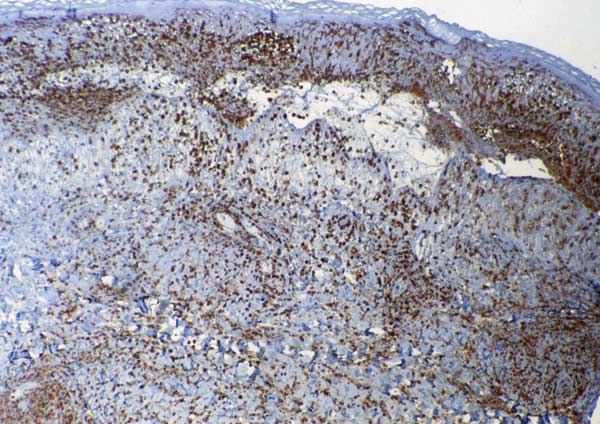
Figure 3. Inoculation eschar from a patient with African tick-bite fever showing numerous dermal inflammatory infiltrates mainly composed of polymorphonuclear leukocytes (immunoperoxidase staining with an anti-CD15 antibody; original magnification ×100).
Page created: November 17, 2011
Page updated: November 17, 2011
Page reviewed: November 17, 2011
The conclusions, findings, and opinions expressed by authors contributing to this journal do not necessarily reflect the official position of the U.S. Department of Health and Human Services, the Public Health Service, the Centers for Disease Control and Prevention, or the authors' affiliated institutions. Use of trade names is for identification only and does not imply endorsement by any of the groups named above.