Volume 13, Number 10—October 2007
Dispatch
Isolation of Bartonella sp. from Sheep Blood
David A. Bemis* and Stephen A. Kania*
Figure 1
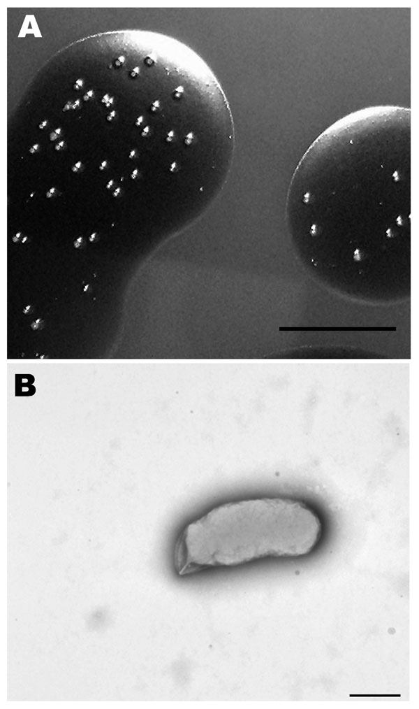
Figure 1. Morphologic analysis of a Bartonella sp. isolated from sheep blood. A) Colonies growing in sheep blood surface biofilm seen in reflected light after 25 days. Scale bar = 10 mm. B) Transmission electron micrograph of a representative cell that was dispersed from a 25-day-old colony and negatively stained with 0.5% potassium phosphotungstic acid. Scale bar = 500 nm
Page created: July 02, 2010
Page updated: July 02, 2010
Page reviewed: July 02, 2010
The conclusions, findings, and opinions expressed by authors contributing to this journal do not necessarily reflect the official position of the U.S. Department of Health and Human Services, the Public Health Service, the Centers for Disease Control and Prevention, or the authors' affiliated institutions. Use of trade names is for identification only and does not imply endorsement by any of the groups named above.