Volume 13, Number 11—November 2007
Dispatch
Rocky Mountain Spotted Fever, Panama
Figure 1

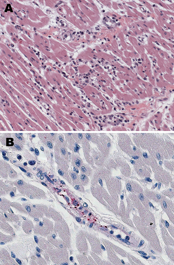
Figure 1. Histologic and immunohistochemical evaluation of heart tissue. A) Lymphohistiocytic inflammatory cell infiltrates in the myocardium (hematoxylin and eosin stain; original magnification ×25). B) Immunohistochemical detection of spotted fever group rickettsiae (red) in perivascular infiltrates of heart (immunoalkaline phosphatase with naphthol-fast red substrate and hematoxylin counterstain; original magnification ×250.
Page created: July 06, 2010
Page updated: July 06, 2010
Page reviewed: July 06, 2010
The conclusions, findings, and opinions expressed by authors contributing to this journal do not necessarily reflect the official position of the U.S. Department of Health and Human Services, the Public Health Service, the Centers for Disease Control and Prevention, or the authors' affiliated institutions. Use of trade names is for identification only and does not imply endorsement by any of the groups named above.