Volume 13, Number 2—February 2007
Letter
Misdiagnosing Melioidosis
Figure
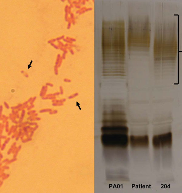
Figure. A) Gram stain of pus from the patient’s submental collection, showing the characteristic safety-pin pattern (arrows) of bipolar staining. B) Sodium dodecyl sulfate–polyacrylamide gel electrophoresis of lipopolysaccharide (LPS) antigens from the patient and Burkholderia pseudomallei reference strain (204), showing different O-repeating units (bracket). A control isolate of Pseudomonas aeruginosa LPS (PA01) is shown for comparison.
1A. J. Brent and R. Handy had clinical responsibility for the patient. R. Handy and P.C. Matthews made the initial microbiologic diagnosis of melioidosis, and T.L. Pitt confirmed the isolate as Burkholderia pseudomallei. T.L. Pitt performed the serology and SDS-PAGE analysis of lipopolysaccharide antigens. All authors contributed to preparation of the manuscript. A.J. Brent is guarantor for the article, had full access to all the clinical and microbiologic data, and had final responsibility for the decision to submit for publication.