Volume 14, Number 8—August 2008
Dispatch
Rickettsia felis in Fleas, Germany
Cite This Article
Citation for Media
Abstract
Among 310 fleas collected from dogs and cats in Germany, Rickettsia felis was detected in all specimens (34) of Archaeopsylla erinacei (hedgehog flea) and in 9% (24/226) of Ctenocephalides felis felis (cat flea). R. helvetica was detected in 1 Ceratophyllus gallinae (hen flea).
Rickettsia felis, the causative agent of the flea-borne spotted fever rickettsiosis, is pathogenic for humans (1–4). Since the first detection of R. felis from midgut epithelial cells of the cat flea, Ctenocephalides felis felis, in 1990 (5), interest in the role of this flea species as its main vector has increased. R. felis has been found in cat fleas on all continents (6). Because R. felis is not lethal for cat fleas and is transmitted transovarially by these fleas (4), C. felis could be a vector and a reservoir of this pathogen. For these reasons, the cat flea was considered the only flea species with a major role in the epidemiology of flea-borne spotted fever rickettsiosis. However, R. felis has been reported in other flea species (4,6–8), and flea-borne spotted fever rickettsiosis is now considered an emerging human infectious disease. We analyzed the presence of R. felis in different flea species collected from naturally infested cats and dogs in different locations in Germany.
Figure
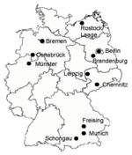
Figure. Locations of the 11 flea-collection study sites in Germany, 2007.
A total of 310 fleas were collected from 49 dogs and 54 cats in 11 widely distributed locations in Germany (Berlin, Munich, Brandenburg, Leipzig, Chemnitz, Rostock/Laage, Bremen, Osnabrück, Münster, Freising, and Schongau) (Figure) in 2007. Specimens collected were recorded and kept at –20°C. Samples were shipped on dry ice to our laboratory, and species identification was performed by using light microscopy and following the determination key of Hopkins and Rothschild (9). Because of infestation variations (1–150 fleas per animal), 3 fleas per animal host were chosen randomly for species differentiation.
Fleas were homogenized individually in 80 μL of phosphate-buffered saline with a RETSCH Tissue Lyser Mixer Mill 300 (QIAGEN, Hilden, Germany) by using 5-mm steel beads. A 100-μL volume of ATL buffer and 20 μL of proteinase K (QIAGEN) were added, and homogenates were incubated at 56°C in an Eppendorf Thermomixer (Eppendorf, Hamburg, Germany) until tissues were completely lysed. DNA was extracted from each flea by using a QIAamp DNA Mini Kit (QIAGEN) according to the manufacturer’s instructions (tissue protocol) and stored at –20°C until used.
PCR amplification of rickettsial DNA was performed by using oligonucleotide primer pairs Rp CS.877p/Rp CS.1258n (10) generated from the rickettsial citrate synthase (gltA) gene. Positive samples were analyzed for a 530-bp portion of the outer membrane protein A (ompA) gene with primer pair Rr 190.70p/Rr 190.602n (10) and for a 765-bp portion of the ompB gene with primer pair 120–1278/120–3599 (11). PCR amplification was accomplished in 50-μL volumes containing 5 μL DNA, 30 μL distilled water, 10 μL 5× Taq buffer (Roche, Mannheim, Germany), 3 μL 25 mmol/L MgCl2 (Roche), 1 μL 10 mmol/L dNTP (Roche), 0.25 μL each primer (100 μmol/L), and 0.5 μL Taq polymerase (5U/mL; Roche). Conditions for the gltA and ompA PCRs were as described by Bertolotti et al. (12). Negative and positive controls were included in all PCRs.
All PCR products were separated by electrophoresis on 1.5% agarose gels at 100 V for 60 min and examined under UV light. Positive samples for both genes were purified by using the QIAquick PCR purification kit (QIAGEN) and sequenced by the MWG Biotech Company (Martinried, Germany). Sequences obtained were compared with those of characterized rickettsia in GenBank by using BLAST analysis (www.ncbi.nlm.nih.gov).
Five species of fleas were identified in the study. The most prevalent species was C. felis (93% of fleas in cats and 78% in dogs) (Table 1). Archaeopsylla erinacei (hedgehog flea), was the second most abundant species, with 26 specimens collected from dogs and 8 from cats. A few specimens of Ctenocephalides canis (dog flea), Pulex irritans (human flea), and Ceratophyllus gallinae (hen flea) were also identified (Table 1). Eight dogs had mixed populations of fleas; 5 had C. felis and A. erinacei, 2 had C. felis and C. gallinae, and 1 had C. felis and C. canis. Mixed populations of fleas were also detected in 3 cats; 2 were infested with C. felis and A. erinacei, and 1 with P. irritans and C. felis.
Thirty-six (25%) of 146 fleas collected from dogs and 24 (15%) of 164 fleas collected from cats were positive for the gltA gene. Positive fleas were found in 6 of 11 sampled locations. Proportions of infected fleas collected from dogs ranged from 25% (Berlin) to 56% (Münster), and proportions of infected fleas collected from cats ranged from 10% (Freising) to 100% (Münster) (Table 2).
Of 60 fleas positive for the gltA gene (for dogs and cats), only 2 were negative for the ompA and ompB genes. Sequencing analysis of the gltA gene for these 2 samples showed that 1 sequence (from C. gallinae) was 99% homologous with part of the Rickettsia helveticagltA gene (AM418450.1) from an Ixodes persulcatus tick isolated in Russia; the other sequence (from C. gallinae) was 94% homologous with the Rickettsia sp. citrate synthase gene (U76908.1). Thus, we report R. helvetica in C. gallinae ticks.
Of the other 58 gltA-positive samples, 2 were positive for the ompA gene in the first round; 56 fleas were positive for the ompB gene. The 2 ompA-positive samples were sequenced, and sequences matched the ompA gene from R. felis (AJ563398.1; 99%–100% similarity). The 56 positive ompB samples were sequenced, and sequences matched with ompB gene from R. felis (CP000053.1; 98%–100% similarity). All hedgehog fleas (34 specimens) collected were infected with R. felis. Moreover, these 34 specimens were collected from 5 locations within a large area from Berlin (northeastern Germany) to Munich (southeastern Germany). Our findings indicate that A. erinacei may play a major role in the transmission of R. felis in Germany. Recent studies reported R. felis in 1 hedgehog flea in Portugal (13) and in 4 hedgehog fleas in Algeria (8).
Our study confirms that C. felis remains the most common flea species infesting cats and dogs in Germany. Nevertheless, only 24 of 266 cat fleas collected were infected with R. felis. Infected cat fleas were found only in 4 of 11 studied sites, in contrast with a recent study in France, where R. felis–infected C. felis were present in all locations studied (14). In the 4 positive sites in Germany, 3 had positive A. erinacei specimens and 1 had positive C. gallinae (Table 2). In the other sites where no positive fleas where found, only C. felis was present either alone or in association with P. irritans and C. canis (Table 2).
Although C. felis seems to be the main vector of R. felis, our findings indicate that A. erinacei may be a vector for human flea-borne rickettsiosis in Germany. Because hedgehogs may act as a reservoir of pathogens (15), further studies will be conducted to investigate the role of hedgehogs and hedgehog fleas in maintenance and transmission of R. felis in Germany.
Dr Gilles is National Institutes of Health project leader at the University of Kentucky in Lexington. His research interests focus on medical entomology, arthropod-borne diseases, vector biology, ecology, and vector control.
References
- Schriefer ME, Sacci JBJr, Dumler JS, Bullen MG, Azad AF. Identification of a novel rickettsial infection in a patient diagnosed with murine typhus.J Clin Microbiol. 1994;32:949–54.PubMedGoogle Scholar
- Zavala-Velazquez JE, Sosa-Ruiz JA, Zavala-Castro J, Jimenez-Delgadillo B, Vado-Solis IE, Sanchez-Elias RA, Rickettsia felis: the etiologic agent of three cases of rickettsiosis in Yucatan.Lancet. 2000;356:1079–80. DOIPubMedGoogle Scholar
- Richter J, Fournier P, Petridou J, Haussinger D, Raoult D. Rickettsia felis infection acquired in Europe and documented by polymerase chain reaction.Emerg Infect Dis. 2002;8:207–8.PubMedGoogle Scholar
- Rolain J-M, Franc M, Davoust B, Raoult D. Molecular detection of Bartonella quintana, B. koehlerae, B. henselae, B. clarridgeiae, Rickettsia felis, and Wolbachia pipientis in cat fleas, France.Emerg Infect Dis. 2003;9:338–42.PubMedGoogle Scholar
- Adams JR, Schmidtmann ET, Azad AF. Infection of colonized cat fleas, Ctenocephalides felis (Bouché), with a rickettsia-like microorganism.Am J Trop Med Hyg. 1990;43:400–9.PubMedGoogle Scholar
- Stevenson HL, Labruna MB, Montenieri JA, Kosoy MY, Gage KL, Walker DH. Detection of Rickettsia felis in a New World flea species, Anomiopsyllus nudata (Siphonaptera: Ctenophthalmidae).J Med Entomol. 2005;42:163–7. DOIPubMedGoogle Scholar
- Jiang J, Soeatmadji DW, Henry KM, Ratiwayanto S, Bangs MJ, Richards AL. Rickettsia felis in Xenopsylla cheopis, Java, Indonesia.Emerg Infect Dis. 2006;12:1281–3.PubMedGoogle Scholar
- Bitam I, Parola P, De La Cruz KD, Matsumoto K, Baziz B, Rolain JM, First molecular detection of Rickettsia felis in fleas from Algeria.Am J Trop Med Hyg. 2006;74:532–5.PubMedGoogle Scholar
- Hopkins GH, Rothschild M. An illustrated catalogue 1 of the Rothschild collection of fleas (Siphonaptera) in the British Museum (Natural History). With keys and short descriptions for the identification of families, genera, species and subspecies of the order. Vol. IV. Ctenophthalmidae, Dino-psyllidae, Doratopsyllidae and Listropsyllidae. London: British Museum (Natural History); 1966.
- Regnery RL, Spruill CL, Plikaytis BD. Genotypic identification of rickettsiae and estimation of intraspecies sequence divergence for portions of two rickettsial genes.J Bacteriol. 1991;173:1576–89.PubMedGoogle Scholar
- Roux V, Raoult D. Phylogenetic analysis of members of the genus Rickettsia using the gene encoding the outer-membrane protein rOmpB (ompB).Int J Syst Evol Microbiol. 2000;50:1449–55.PubMedGoogle Scholar
- Bertolotti L, Tomassone L, Tramuta C, Greco E, Amore G, Ambrogi C, Borrelia lusitaniae and spotted fever group rickettsiae in Ixodes ricinus (Acari: Ixodidae) in Tuscany, Central Italy.J Med Entomol. 2006;43:159–65. DOIPubMedGoogle Scholar
- de Sousa R, Edouard-Fournier P, Santos-Silva M, Amaro F, Bacellar F, Raoult D. Molecular detection of Rickettsia felis, Rickettsia typhi and two genotypes closely related to Bartonella elizabethae.Am J Trop Med Hyg. 2006;75:727–31.PubMedGoogle Scholar
- Gilles J, Just FT, Silaghi C, Pradel I, Lengauer H, Hellmann K, Rickettsia felis in fleas, France[letter]. Emerg Infect Dis. 2008;14:684–6.PubMedGoogle Scholar
Figure
Tables
Cite This ArticleTable of Contents – Volume 14, Number 8—August 2008
| EID Search Options |
|---|
|
|
|
|
|
|
Please use the form below to submit correspondence to the authors or contact them at the following address:
Jérémie Gilles, Department of Entomology, University of Kentucky, S225, Agricultural Science Center North, Lexington, KY 40506, USA;
Top