Volume 14, Number 8—August 2008
Dispatch
Endemic Circulation of European Bat Lyssavirus Type 1 in Serotine Bats, Spain
Sonia Vázquez-Morón*† , Javier Juste‡, Carlos Ibáñez‡, Eduardo Ruiz-Villamor§, Ana Avellón*, Manuel Vera*, and Juan E. Echevarría*†
Figure 2
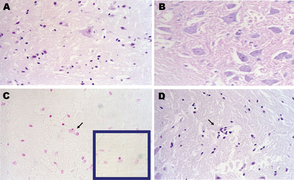
Figure 2. Pathologic images obtained from a carcass of Eptesicus isabellinus. The bat was captured while flying but died during handling. Brain specimen was positive for lyssavirus antigens by immunofluorescence and for European bat lyssavirus 1 RNA by reverse transcription–PCR. A) Neural degeneration in brain by hematoxylin and eosin stain (H&E); magnification ×400. B) Negative Seller stain in spinal cord indicating the absence of Negri bodies; magnification ×400. C) Positive Feulgen reaction in brain, glial cell neurophagia; magnification ×200. D) Focal proliferation of glial cells by H&E; magnification ×100.
Page created: July 12, 2010
Page updated: July 12, 2010
Page reviewed: July 12, 2010
The conclusions, findings, and opinions expressed by authors contributing to this journal do not necessarily reflect the official position of the U.S. Department of Health and Human Services, the Public Health Service, the Centers for Disease Control and Prevention, or the authors' affiliated institutions. Use of trade names is for identification only and does not imply endorsement by any of the groups named above.