Volume 15, Number 12—December 2009
Dispatch
Molecular Model of Prion Transmission to Humans
Figure 1
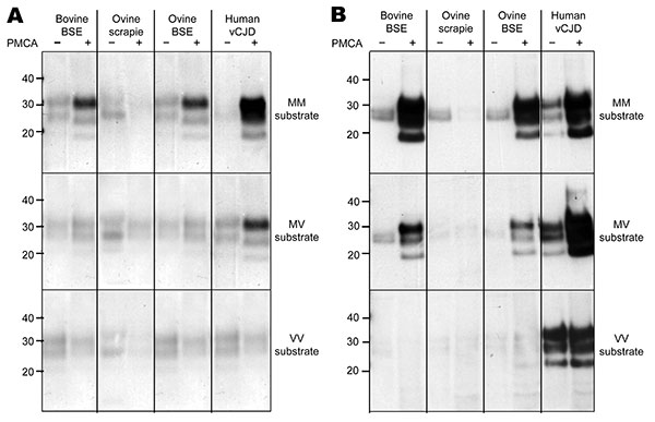
Figure 1. Amplification of PrPd by PMCA from bovine BSE, ovine scrapie, experimental ovine BSE, and human vCJD brain homogenates in substrate homogenates prepared from humanized transgenic mouse brain tissue expressing PrP of each human prion protein gene codon 129 (PRNP-129) genotype. A) Amplification of each PrPd type, as determined by Western blotting using MAb 6H4 to detect PrPres after limited proteinase K digestion, in a PRNP-129MM substrate (top panel, 3-min exposure), a PRNP-129MV substrate (middle panel, 3-min exposure), and a PRNP-129VV substrate (bottom panel, 3-min exposure). B) Amplification of each PrPd type, as determined by Western blotting using MAb 3F4 to detect PrPres derived from human PrP after limited proteinase K digestion, in a PRNP-129MM substrate (top panel, 30-s exposure), a PRNP-129MV substrate (middle panel, 3-min exposure), and a PRNP-129VV substrate (bottom panel, 10-min exposure). Limited proteinase K digestion and Western blotting were conducted out as previously described (11). MAb 6H4 (Prionics, Schlieren-Zurich, Switzerland) and MAb 3F4 (Dako, Ely, Cambridgeshire, UK) were used at a final concentration of 50 ng/mL. PrPd, disease-associated prion protein; PMCA, protein misfolding cyclic amplification; BSE, bovine spongiform encephalopathy; vCJD, variant Creutzfeldt-Jakob disease; MAb, monoclonal antibody; PrPres, protease-resistant prion protein; MM, methionine homozygous; MV, methionine/valine heterozygous; VV, valine homozygous. Values on the left are in kilodaltons.
References
- Collinge J, Clark AR. A general model of prion strains and their pathogenicity. Science. 2007;318:930–6. DOIPubMedGoogle Scholar
- Will RG, Ironside JW, Zeidler M, Cousens SN, Estibeiro K, Alperovitch A, Lancet. 1996;347:921–5. DOIPubMedGoogle Scholar
- Collinge J, Sidle KC, Meads J, Ironside J, Hill AF. Molecular analysis of prion strain variation and the aetiology of ‘new variant’ CJD. Nature. 1996;383:685–90. DOIPubMedGoogle Scholar
- Head MW, Bunn TJ, Bishop MT, McLoughlin V, Lowrie S, McKimmie J, Prion protein heterogeneity in sporadic but not variant Creutzfeldt-Jakob disease: UK cases 1991–2002. Ann Neurol. 2004;55:851–9. DOIPubMedGoogle Scholar
- Eloit M, Adjou K, Coulpier M, Fontaine JJ, Hamel R, Lilin T, BSE agent signatures in a goat. Vet Rec. 2005;156:523–4.PubMedGoogle Scholar
- Supattapone S. Prion protein conversion in vitro. J Mol Med. 2004;82:348–56. DOIPubMedGoogle Scholar
- Saborio GP, Permanne B, Soto C. Sensitive detection of pathological prion protein by cyclic amplification of protein misfolding. Nature. 2001;411:810–3. DOIPubMedGoogle Scholar
- Jones M, Peden AH, Prowse CV, Groner A, Manson JC, Turner ML, In vitro amplification and detection of variant Creutzfeldt-Jakob disease PrPSc. J Pathol. 2007;213:21–6. DOIPubMedGoogle Scholar
- Yull HM, Ritchie DL, Langeveld JP, van Zijderveld FG, Bruce ME, Ironside JW, Detection of type 1 prion protein in variant Creutzfeldt-Jakob disease. Am J Pathol. 2006;168:151–7. DOIPubMedGoogle Scholar
- Jeffrey M, Martin S, Thomson JR, Dingwall WS, Begara-McGorum I, Gonzalez L. Onset and distribution of tissue prp accumulation in scrapie-affected Suffolk sheep as demonstrated by sequential necropsies and tonsillar biopsies. J Comp Pathol. 2001;125:48–57. DOIPubMedGoogle Scholar
- Gonzalez L, Chianini F, Martin S, Siso S, Gibbard L, Reid HW, Comparative titration of experiment ovine BSE infectivity in sheep and mice. J Gen Virol. 2007;88:714–7. DOIPubMedGoogle Scholar
- Bishop MT, Hart P, Aitchison L, Baybutt HN, Plinston C, Thomson V, Predicting susceptibility and incubation time of human-to-human transmission of vCJD. Lancet Neurol. 2006;5:393–8. DOIPubMedGoogle Scholar
- Manson JC, Cancellotti E, Hart P, Bishop MT, Barron RM. The transmissible spongiform encephalopathies: emerging and declining epidemics. Biochem Soc Trans. 2006;34:1155–8. DOIPubMedGoogle Scholar