Volume 15, Number 8—August 2009
Research
Serologic Evidence of Frequent Human Infection with WU and KI Polyomaviruses
Figure 4

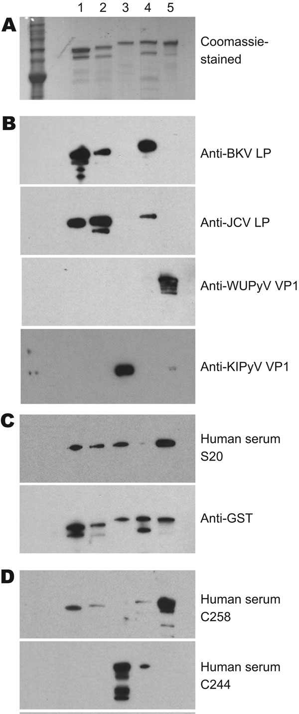
Figure 4. Results of patient serum sample Western blotting for polyomaviruses. A) Coomassie blue–stained image showing 5 types of purified glutathione S-transferase (GST)–tagged viral protein 1 (VP1) in a sodium dodecyl sulfate–polyacrylamide gel. Lane 1, GST-BKV VP1; lane 2, GST-JCV VP1; lane 3, GST–KI polyomavirus (KIPyV) VP1; lane 4, GST-SV40 VP1; lane 5, GST–WU polyomavirus (WUPyV) VP1. B) Western blot results using control rabbit antiserum against BK virus-like particles (BKVLP), JC virus-like particles (JCVLP), WUPyV VP1, or KIPyV VP1 as primary antibody. C) Western blot results for serum that was positive (S20) for WU polyomavirus and KI polyomavirus by ELISA. Antibody against GST was used as a loading control. D) Western blot result for serum that was ELISA positive for WU (C258) and KI (C244).