Volume 16, Number 10—October 2010
Letter
Dictyostelium polycephalum Infection of Human Cornea
Ashok Kumar Reddy1 , Praveen Kumar Balne, Prashant Garg, Virender Singh Sangwan, Madhusmita Das, Pravin V. Krishna, Bhupesh Bagga, and Geeta K. Vemuganti
Figure
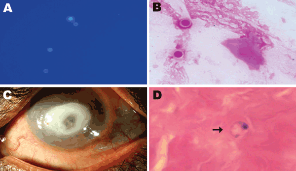
Figure. A) Spherical cysts of Dictyostelium spp. in potassium hydroxide (calcoflour white stain; original magnification ×40) preparation. B) Spherical double wall cysts of Dictyostelium spp. (Gram stain; original magnification ×100). C) Cornea of the patient’s left eye, showing a ring-shaped central infiltrate and central thinning. D) Corneal button showing Dictyostelium spp. cysts (arrow; hematoxylin and eosin stain; original magnification ×100).
1Current affiliation: GHR Micro Diagnostics,Hyderabad, India.
Page created: September 08, 2011
Page updated: September 08, 2011
Page reviewed: September 08, 2011
The conclusions, findings, and opinions expressed by authors contributing to this journal do not necessarily reflect the official position of the U.S. Department of Health and Human Services, the Public Health Service, the Centers for Disease Control and Prevention, or the authors' affiliated institutions. Use of trade names is for identification only and does not imply endorsement by any of the groups named above.