Research
Influenza A (H5N1) Viruses from Pigs, Indonesia [PDF - 722 KB - 9 pages]
Pigs have long been considered potential intermediate hosts in which avian influenza viruses can adapt to humans. To determine whether this potential exists for pigs in Indonesia, we conducted surveillance during 2005–2009. We found that 52 pigs in 4 provinces were infected during 2005–2007 but not 2008–2009. Phylogenetic analysis showed that the viruses had been introduced into the pig population in Indonesia on at least 3 occasions. One isolate had acquired the ability to recognize a human-type receptor. No infected pig had influenza-like symptoms, indicating that influenza A (H5N1) viruses can replicate undetected for prolonged periods, facilitating avian virus adaptation to mammalian hosts. Our data suggest that pigs are at risk for infection during outbreaks of influenza virus A (H5N1) and can serve as intermediate hosts in which this avian virus can adapt to mammals.
| EID | Nidom CA, Takano R, Yamada S, Sakai-Tagawa Y, Daulay S, Aswadi D, et al. Influenza A (H5N1) Viruses from Pigs, Indonesia. Emerg Infect Dis. 2010;16(10):1515-1523. https://doi.org/10.3201/eid1610.100508 |
|---|---|
| AMA | Nidom CA, Takano R, Yamada S, et al. Influenza A (H5N1) Viruses from Pigs, Indonesia. Emerging Infectious Diseases. 2010;16(10):1515-1523. doi:10.3201/eid1610.100508. |
| APA | Nidom, C. A., Takano, R., Yamada, S., Sakai-Tagawa, Y., Daulay, S., Aswadi, D....Kawaoka, Y. (2010). Influenza A (H5N1) Viruses from Pigs, Indonesia. Emerging Infectious Diseases, 16(10), 1515-1523. https://doi.org/10.3201/eid1610.100508. |
Mobile Phone–based Infectious Disease Surveillance System, Sri Lanka [PDF - 297 KB - 8 pages]
Because many infectious diseases are emerging in animals in low-income and middle-income countries, surveillance of animal health in these areas may be needed for forecasting disease risks to humans. We present an overview of a mobile phone–based frontline surveillance system developed and implemented in Sri Lanka. Field veterinarians reported animal health information by using mobile phones. Submissions increased steadily over 9 months, with ≈4,000 interactions between field veterinarians and reports on the animal population received by the system. Development of human resources and increased communication between local stakeholders (groups and persons whose actions are affected by emerging infectious diseases and animal health) were instrumental for successful implementation. The primary lesson learned was that mobile phone–based surveillance of animal populations is acceptable and feasible in lower-resource settings. However, any system implementation plan must consider the time needed to garner support for novel surveillance methods among users and stakeholders.
| EID | Robertson C, Sawford K, Daniel SL, Nelson TA, Stephen C. Mobile Phone–based Infectious Disease Surveillance System, Sri Lanka. Emerg Infect Dis. 2010;16(10):1524-1531. https://doi.org/10.3201/eid1610.100249 |
|---|---|
| AMA | Robertson C, Sawford K, Daniel SL, et al. Mobile Phone–based Infectious Disease Surveillance System, Sri Lanka. Emerging Infectious Diseases. 2010;16(10):1524-1531. doi:10.3201/eid1610.100249. |
| APA | Robertson, C., Sawford, K., Daniel, S. L., Nelson, T. A., & Stephen, C. (2010). Mobile Phone–based Infectious Disease Surveillance System, Sri Lanka. Emerging Infectious Diseases, 16(10), 1524-1531. https://doi.org/10.3201/eid1610.100249. |
Oral Fluid Testing during 10 Years of Rubella Elimination, England and Wales [PDF - 324 KB - 7 pages]
Surveillance of rubella in England and Wales has included immunoglobulin M testing of oral (crevicular) fluid from reported case-patients since 1994. The need for laboratory confirmation to monitor rubella elimination is emphasized by poor sensitivity (51%, 95% confidence interval 48.9%–54.0%) and specificity (55%, 95% confidence interval 53.7%–55.6%) of the clinical case definition. During 1999–2008, oral fluid from 11,709 (84%) of 13,952 reported case-patients was tested; 143 (1.0%) cases were confirmed and 11,566 (99%) were discarded (annual investigation and discard rate of clinically suspected rubella cases was 2,208/100,000 population). Incidence of confirmed rubella increased from 0.50 to 0.77/1 million population when oral fluid testing was included. Oral fluid tests confirmed that cases were more likely to be in older, unvaccinated men. Testing of oral fluid has improved ascertainment of confirmed rubella in children and men and provided additional information for assessing UK progress toward the World Health Organization elimination goal.
| EID | Manikkavasagan G, Bukasa A, Brown KE, Cohen BJ, Ramsay ME. Oral Fluid Testing during 10 Years of Rubella Elimination, England and Wales. Emerg Infect Dis. 2010;16(10):1532-1538. https://doi.org/10.3201/eid1610.100560 |
|---|---|
| AMA | Manikkavasagan G, Bukasa A, Brown KE, et al. Oral Fluid Testing during 10 Years of Rubella Elimination, England and Wales. Emerging Infectious Diseases. 2010;16(10):1532-1538. doi:10.3201/eid1610.100560. |
| APA | Manikkavasagan, G., Bukasa, A., Brown, K. E., Cohen, B. J., & Ramsay, M. E. (2010). Oral Fluid Testing during 10 Years of Rubella Elimination, England and Wales. Emerging Infectious Diseases, 16(10), 1532-1538. https://doi.org/10.3201/eid1610.100560. |
Human Monkeypox Outbreak Caused by Novel Virus Belonging to Congo Basin Clade, Sudan, 2005 [PDF - 316 KB - 7 pages]
To determine the outbreak source of monkeypox virus (MPXV) infections in Unity State, Sudan, in November 2005, we conducted a retrospective investigation. MPXV was identified in a sub-Sahelian savannah environment. Three case notification categories were used: suspected, probable, and confirmed. Molecular, virologic, and serologic assays were used to test blood specimens, vesicular swabs, and crust specimens obtained from symptomatic and recovering persons. Ten laboratory-confirmed cases and 9 probable cases of MPXV were reported during September–December 2005; no deaths occurred. Human-to-human transmission up to 5 generations was described. Our investigation could not fully determine the source of the outbreak. Preliminary data indicate that the MPXV strain isolated during this outbreak was a novel virus belonging to the Congo Basin clade. Our results indicate that MPXV should be considered endemic to the wetland areas of Unity State. This finding will enhance understanding of the ecologic niche for this virus.
| EID | Formenty P, Muntasir MO, Damon IK, Chowdhary V, Opoka ML, Monimart C, et al. Human Monkeypox Outbreak Caused by Novel Virus Belonging to Congo Basin Clade, Sudan, 2005. Emerg Infect Dis. 2010;16(10):1539-1545. https://doi.org/10.3201/eid1610.100713 |
|---|---|
| AMA | Formenty P, Muntasir MO, Damon IK, et al. Human Monkeypox Outbreak Caused by Novel Virus Belonging to Congo Basin Clade, Sudan, 2005. Emerging Infectious Diseases. 2010;16(10):1539-1545. doi:10.3201/eid1610.100713. |
| APA | Formenty, P., Muntasir, M. O., Damon, I. K., Chowdhary, V., Opoka, M. L., Monimart, C....Abdalla, M. S. (2010). Human Monkeypox Outbreak Caused by Novel Virus Belonging to Congo Basin Clade, Sudan, 2005. Emerging Infectious Diseases, 16(10), 1539-1545. https://doi.org/10.3201/eid1610.100713. |
Therapeutic Drug Monitoring for Slow Response to Tuberculosis Treatment in a State Control Program, Virginia, USA [PDF - 316 KB - 8 pages]
Therapeutic drug monitoring may be useful in tuberculosis management, but programmatic implementation is understudied. We performed a retrospective cohort study to determine prevalence of lower than expected levels of isoniazid, rifampin, ethambutol, and pyrazinamide measured at time of estimated peak serum concentration. Patients were tested for serum concentration at 2 hours after medication administration. When patients were tested, 22 had concentrations lower than expected range for rifampin, 23 of 39 patients had low levels of isoniazid, and 8 of 26 patients had low levels of ethambutol; all 20 patients tested for pyrazinamide were within expected range. Over 26 months, 42 patients met criteria for slow response. Diabetes was associated with slow response (p<0.001), and persons with diabetes were more likely than persons without diabetes to have low rifampin levels (p = 0.03). Dosage adjustment of rifampin was more likely to elevate serum concentration to the target range than adjustment of isoniazid given in daily doses (p = 0.01).
| EID | Heysell SK, Moore JL, Keller SJ, Houpt ER. Therapeutic Drug Monitoring for Slow Response to Tuberculosis Treatment in a State Control Program, Virginia, USA. Emerg Infect Dis. 2010;16(10):1546-1553. https://doi.org/10.3201/eid1610.100374 |
|---|---|
| AMA | Heysell SK, Moore JL, Keller SJ, et al. Therapeutic Drug Monitoring for Slow Response to Tuberculosis Treatment in a State Control Program, Virginia, USA. Emerging Infectious Diseases. 2010;16(10):1546-1553. doi:10.3201/eid1610.100374. |
| APA | Heysell, S. K., Moore, J. L., Keller, S. J., & Houpt, E. R. (2010). Therapeutic Drug Monitoring for Slow Response to Tuberculosis Treatment in a State Control Program, Virginia, USA. Emerging Infectious Diseases, 16(10), 1546-1553. https://doi.org/10.3201/eid1610.100374. |
Risk Factors for Pandemic (H1N1) 2009 Virus Seroconversion among Hospital Staff, Singapore [PDF - 222 KB - 8 pages]
We describe incidence and risk factors for pandemic (H1N1) 2009 virus infection in healthcare personnel during the June–September 2009 epidemic in Singapore. Personnel contributed 3 serologic samples during June–October 2009, with seroconversion defined as a >4-fold increase in hemagglutination inhibition titers to pandemic (H1N1) 2009. Of 531 participants, 35 showed evidence of seroconversion. Seroconversion rates were highest in nurses (28/290) and lowest in allied health staff (2/116). Significant risk factors on multivariate analysis were being a nurse (adjusted odds ratio [aOR] 4.5, 95% confidence interval [CI] 1.0–19.6) and working in pandemic (H1N1) 2009 isolation wards (aOR 4.5, 95% CI 1.3–15.6). Contact with pandemic (H1N1) 2009–infected colleagues (aOR 2.5, 95% CI 0.9–6.6) and larger household size (aOR 1.2, 95% CI 1.0–1.4) were of borderline significance. Our study suggests that seroconversion was associated with occupational and nonoccupational risk factors.
| EID | Chen MI, Lee VJ, Barr I, Lin C, Goh R, Lee C, et al. Risk Factors for Pandemic (H1N1) 2009 Virus Seroconversion among Hospital Staff, Singapore. Emerg Infect Dis. 2010;16(10):1554-1561. https://doi.org/10.3201/eid1610.100516 |
|---|---|
| AMA | Chen MI, Lee VJ, Barr I, et al. Risk Factors for Pandemic (H1N1) 2009 Virus Seroconversion among Hospital Staff, Singapore. Emerging Infectious Diseases. 2010;16(10):1554-1561. doi:10.3201/eid1610.100516. |
| APA | Chen, M. I., Lee, V. J., Barr, I., Lin, C., Goh, R., Lee, C....Leo, Y. (2010). Risk Factors for Pandemic (H1N1) 2009 Virus Seroconversion among Hospital Staff, Singapore. Emerging Infectious Diseases, 16(10), 1554-1561. https://doi.org/10.3201/eid1610.100516. |
Effectiveness of Personal Protective Equipment and Oseltamivir Prophylaxis during Avian Influenza A (H7N7) Epidemic, the Netherlands, 2003 [PDF - 215 KB - 7 pages]
We analyzed the effectiveness of personal protective equipment and oseltamivir use during the 2003 avian influenza A (H7N7) epidemic in the Netherlands by linking databases containing information about farm visits, human infections, and use of oseltamivir and personal protective equipment. Using a stringent case definition, based on self-reported conjunctivitis combined with a positive hemagglutination-inhibition assay, we found that prophylactic treatment with oseltamivir significantly reduced the risk for infection per farm visit from 0.145 (95% confidence interval [CI] 0.078–0.233) to 0.031 (95% CI 0.008–0.073). The protective effect was ≈79% (95% CI 40%–97%). These results are comparable with the reported effect of prophylactic treatment with oseltamivir on human seasonal influenza. No significant protective effect was found for use of respirators or safety glasses, possibly because of limitations of the data.
| EID | te Beest DE, van Boven M, Bos M, Stegeman A, Koopmans M. Effectiveness of Personal Protective Equipment and Oseltamivir Prophylaxis during Avian Influenza A (H7N7) Epidemic, the Netherlands, 2003. Emerg Infect Dis. 2010;16(10):1562-1568. https://doi.org/10.3201/eid1610.091412 |
|---|---|
| AMA | te Beest DE, van Boven M, Bos M, et al. Effectiveness of Personal Protective Equipment and Oseltamivir Prophylaxis during Avian Influenza A (H7N7) Epidemic, the Netherlands, 2003. Emerging Infectious Diseases. 2010;16(10):1562-1568. doi:10.3201/eid1610.091412. |
| APA | te Beest, D. E., van Boven, M., Bos, M., Stegeman, A., & Koopmans, M. (2010). Effectiveness of Personal Protective Equipment and Oseltamivir Prophylaxis during Avian Influenza A (H7N7) Epidemic, the Netherlands, 2003. Emerging Infectious Diseases, 16(10), 1562-1568. https://doi.org/10.3201/eid1610.091412. |
Bloodstream infections (BSIs) are a major cause of illness in HIV-infected persons. To evaluate prevalence of and risk factors for BSIs in 2,009 HIV-infected outpatients in Cambodia, Thailand, and Vietnam, we performed a single Myco/F Lytic blood culture. Fifty-eight (2.9%) had a clinically significant BSI (i.e., a blood culture positive for an organism known to be a pathogen). Mycobacterium tuberculosis accounted for 31 (54%) of all BSIs, followed by fungi (13 [22%]) and bacteria (9 [16%]). Of patients for whom data were recorded about antiretroviral therapy, 0 of 119 who had received antiretroviral therapy for >14 days had a BSI, compared with 3% of 1,801 patients who had not. In multivariate analysis, factors consistently associated with BSI were fever, low CD4+ T-lymphocyte count, abnormalities on chest radiograph, and signs or symptoms of abdominal illness. For HIV-infected outpatients with these risk factors, clinicians should place their highest priority on diagnosing tuberculosis.
| EID | Varma JK, McCarthy KD, Tasaneeyapan T, Monkongdee P, Kimerling ME, Buntheoun E, et al. Bloodstream Infections among HIV-Infected Outpatients, Southeast Asia. Emerg Infect Dis. 2010;16(10):1569-1575. https://doi.org/10.3201/eid1610.091686 |
|---|---|
| AMA | Varma JK, McCarthy KD, Tasaneeyapan T, et al. Bloodstream Infections among HIV-Infected Outpatients, Southeast Asia. Emerging Infectious Diseases. 2010;16(10):1569-1575. doi:10.3201/eid1610.091686. |
| APA | Varma, J. K., McCarthy, K. D., Tasaneeyapan, T., Monkongdee, P., Kimerling, M. E., Buntheoun, E....Cain, K. P. (2010). Bloodstream Infections among HIV-Infected Outpatients, Southeast Asia. Emerging Infectious Diseases, 16(10), 1569-1575. https://doi.org/10.3201/eid1610.091686. |
Nontuberculous mycobacteria (NTM) disease is a notifiable condition in Queensland, Australia. Mycobacterial isolates that require species identification are forwarded to the Queensland Mycobacterial Reference Laboratory, providing a central opportunity to capture statewide data on the epidemiology of NTM disease. We compared isolates obtained in 1999 and 2005 and used data from the Queensland notification scheme to report the clinical relevance of these isolates. The incidence of notified cases of clinically significant pulmonary disease rose from 2.2 (1999) to 3.2 (2005) per 100,000 population. The pattern of disease has changed from predominantly cavitary disease in middle-aged men who smoke to fibronodular disease in elderly women. Mycobacterium intracellulare is the main pathogen associated with the increase in isolates speciated in Queensland.
| EID | Thomson RM. Changing Epidemiology of Pulmonary Nontuberculous Mycobacteria Infections. Emerg Infect Dis. 2010;16(10):1576-1583. https://doi.org/10.3201/eid1610.091201 |
|---|---|
| AMA | Thomson RM. Changing Epidemiology of Pulmonary Nontuberculous Mycobacteria Infections. Emerging Infectious Diseases. 2010;16(10):1576-1583. doi:10.3201/eid1610.091201. |
| APA | Thomson, R. M. (2010). Changing Epidemiology of Pulmonary Nontuberculous Mycobacteria Infections. Emerging Infectious Diseases, 16(10), 1576-1583. https://doi.org/10.3201/eid1610.091201. |
Dispatches
Mortality Rate Patterns for Hemorrhagic Fever with Renal Syndrome Caused by Puumala Virus [PDF - 200 KB - 3 pages]
To investigate nephropathia epidemica in Sweden during 1997–2007, we determined case-fatality rates for 5,282 patients with this disease. Overall, 0.4% died of acute nephropathia epidemica <3 months after diagnosis. Case-fatality rates increased with age. Only women showed an increased case-fatality rate during the first year after diagnosis.
| EID | Hjertqvist M, Klein SL, Ahlm C, Klingström J. Mortality Rate Patterns for Hemorrhagic Fever with Renal Syndrome Caused by Puumala Virus. Emerg Infect Dis. 2010;16(10):1584-1586. https://doi.org/10.3201/eid1610.100242 |
|---|---|
| AMA | Hjertqvist M, Klein SL, Ahlm C, et al. Mortality Rate Patterns for Hemorrhagic Fever with Renal Syndrome Caused by Puumala Virus. Emerging Infectious Diseases. 2010;16(10):1584-1586. doi:10.3201/eid1610.100242. |
| APA | Hjertqvist, M., Klein, S. L., Ahlm, C., & Klingström, J. (2010). Mortality Rate Patterns for Hemorrhagic Fever with Renal Syndrome Caused by Puumala Virus. Emerging Infectious Diseases, 16(10), 1584-1586. https://doi.org/10.3201/eid1610.100242. |
Pandemic (H1N1) 2009 Virus on Commercial Swine Farm, Thailand [PDF - 232 KB - 4 pages]
A swine influenza outbreak occurred on a commercial pig farm in Thailand. Outbreak investigation indicated that pigs were co-infected with pandemic (H1N1) 2009 virus and seasonal influenza (H1N1) viruses. No evidence of gene reassortment or pig-to-human transmission of pandemic (H1N1) 2009 virus was found during the outbreak.
| EID | Sreta D, Tantawet S, Ayudhya SN, Thontiravong A, Wongphatcharachai M, Lapkuntod J, et al. Pandemic (H1N1) 2009 Virus on Commercial Swine Farm, Thailand. Emerg Infect Dis. 2010;16(10):1587-1590. https://doi.org/10.3201/eid1610.100665 |
|---|---|
| AMA | Sreta D, Tantawet S, Ayudhya SN, et al. Pandemic (H1N1) 2009 Virus on Commercial Swine Farm, Thailand. Emerging Infectious Diseases. 2010;16(10):1587-1590. doi:10.3201/eid1610.100665. |
| APA | Sreta, D., Tantawet, S., Ayudhya, S. N., Thontiravong, A., Wongphatcharachai, M., Lapkuntod, J....Kitikoon, P. (2010). Pandemic (H1N1) 2009 Virus on Commercial Swine Farm, Thailand. Emerging Infectious Diseases, 16(10), 1587-1590. https://doi.org/10.3201/eid1610.100665. |
Toxoplasma gondii Oocyst–specific Antibodies and Source of Infection [PDF - 167 KB - 3 pages]
Infection source can determine cost-effective public health interventions. To quantify risk of acquiring Toxoplasma gondii from environmental sources versus from meat, we examined serum from pregnant women in Chile. Because 43% had oocyst-specific antibodies, we conclude that contaminated meat remains the primary source of infection but that environmental sources also pose substantial risk.
| EID | Muñoz-Zanzi CA, Fry P, Lesina B, Hill D. Toxoplasma gondii Oocyst–specific Antibodies and Source of Infection. Emerg Infect Dis. 2010;16(10):1591-1593. https://doi.org/10.3201/eid1610.091674 |
|---|---|
| AMA | Muñoz-Zanzi CA, Fry P, Lesina B, et al. Toxoplasma gondii Oocyst–specific Antibodies and Source of Infection. Emerging Infectious Diseases. 2010;16(10):1591-1593. doi:10.3201/eid1610.091674. |
| APA | Muñoz-Zanzi, C. A., Fry, P., Lesina, B., & Hill, D. (2010). Toxoplasma gondii Oocyst–specific Antibodies and Source of Infection. Emerging Infectious Diseases, 16(10), 1591-1593. https://doi.org/10.3201/eid1610.091674. |
Predicting Need for Hospitalization of Patients with Pandemic (H1N1) 2009, Chicago, Illinois, USA [PDF - 195 KB - 4 pages]
In the absence of established guidelines for hospitalization of patients with pandemic (H1N1) 2009, we studied emergency department patients to identify clinical parameters that predict need for hospitalization. Independent predictors of hospitalization include multiple high-risk medical conditions, dyspnea, and hypoxia. These findings are easily applicable, with a 79% positive predictive value for hospitalization.
| EID | Vasoo S, Singh K, Trenholme GM. Predicting Need for Hospitalization of Patients with Pandemic (H1N1) 2009, Chicago, Illinois, USA. Emerg Infect Dis. 2010;16(10):1594-1597. https://doi.org/10.3201/eid1610.091889 |
|---|---|
| AMA | Vasoo S, Singh K, Trenholme GM. Predicting Need for Hospitalization of Patients with Pandemic (H1N1) 2009, Chicago, Illinois, USA. Emerging Infectious Diseases. 2010;16(10):1594-1597. doi:10.3201/eid1610.091889. |
| APA | Vasoo, S., Singh, K., & Trenholme, G. M. (2010). Predicting Need for Hospitalization of Patients with Pandemic (H1N1) 2009, Chicago, Illinois, USA. Emerging Infectious Diseases, 16(10), 1594-1597. https://doi.org/10.3201/eid1610.091889. |
Imported Lassa Fever, Pennsylvania, USA, 2010 [PDF - 143 KB - 3 pages]
We report a case of Lassa fever in a US traveler who visited rural Liberia, became ill while in country, sought medical care upon return to the United States, and subsequently had his illness laboratory confirmed. The patient recovered with supportive therapy. No secondary cases occurred.
| EID | Amorosa V, MacNeil A, McConnell R, Patel A, Dillon KE, Hamilton K, et al. Imported Lassa Fever, Pennsylvania, USA, 2010. Emerg Infect Dis. 2010;16(10):1598-1600. https://doi.org/10.3201/eid1610.100774 |
|---|---|
| AMA | Amorosa V, MacNeil A, McConnell R, et al. Imported Lassa Fever, Pennsylvania, USA, 2010. Emerging Infectious Diseases. 2010;16(10):1598-1600. doi:10.3201/eid1610.100774. |
| APA | Amorosa, V., MacNeil, A., McConnell, R., Patel, A., Dillon, K. E., Hamilton, K....Nichol, S. T. (2010). Imported Lassa Fever, Pennsylvania, USA, 2010. Emerging Infectious Diseases, 16(10), 1598-1600. https://doi.org/10.3201/eid1610.100774. |
Type 2 Diabetes Mellitus and Increased Risk for Malaria Infection [PDF - 164 KB - 4 pages]
A case–control study of 1,466 urban adults in Ghana found that patients with type 2 diabetes mellitus had a 46% increased risk for infection with Plasmodium falciparum. Increase in diabetes mellitus prevalence may put more persons at risk for malaria infection.
| EID | Danquah I, Bedu-Addo G, Mockenhaupt FP. Type 2 Diabetes Mellitus and Increased Risk for Malaria Infection. Emerg Infect Dis. 2010;16(10):1601-1604. https://doi.org/10.3201/eid1610.100399 |
|---|---|
| AMA | Danquah I, Bedu-Addo G, Mockenhaupt FP. Type 2 Diabetes Mellitus and Increased Risk for Malaria Infection. Emerging Infectious Diseases. 2010;16(10):1601-1604. doi:10.3201/eid1610.100399. |
| APA | Danquah, I., Bedu-Addo, G., & Mockenhaupt, F. P. (2010). Type 2 Diabetes Mellitus and Increased Risk for Malaria Infection. Emerging Infectious Diseases, 16(10), 1601-1604. https://doi.org/10.3201/eid1610.100399. |
Epidemiology of Human Parvovirus 4 Infection in Sub-Saharan Africa [PDF - 157 KB - 3 pages]
Human parvovirus 4 infections are primarily associated with parenteral exposure in western countries. By ELISA, we demonstrate frequent seropositivity for antibody to parvovirus 4 viral protein 2 among adult populations throughout sub-Saharan Africa (Burkina Faso, 37%; Cameroon, 25%; Democratic Republic of the Congo, 35%; South Africa, 20%), which implies existence of alternative transmission routes.
| EID | Sharp CP, Vermeulen M, Nébié YK, Djoko CF, LeBreton M, Tamoufe U, et al. Epidemiology of Human Parvovirus 4 Infection in Sub-Saharan Africa. Emerg Infect Dis. 2010;16(10):1605-1607. https://doi.org/10.3201/eid1610.101001 |
|---|---|
| AMA | Sharp CP, Vermeulen M, Nébié YK, et al. Epidemiology of Human Parvovirus 4 Infection in Sub-Saharan Africa. Emerging Infectious Diseases. 2010;16(10):1605-1607. doi:10.3201/eid1610.101001. |
| APA | Sharp, C. P., Vermeulen, M., Nébié, Y. K., Djoko, C. F., LeBreton, M., Tamoufe, U....Simmonds, P. (2010). Epidemiology of Human Parvovirus 4 Infection in Sub-Saharan Africa. Emerging Infectious Diseases, 16(10), 1605-1607. https://doi.org/10.3201/eid1610.101001. |
Artesunate Misuse and Plasmodium falciparum Malaria in Traveler Returning from Africa [PDF - 219 KB - 3 pages]
Plasmodium falciparum malaria developed in an African-born traveler who returned to Canada after visiting Nigeria. While there, she took artesunate prophylactically. Isolates had an elevated 50% inhibitory concentration to artemisinin, artesunate, and artemether, compared with that of other African isolates. Inappropriate use of artemisinin derivatives can reduce P. falciparum susceptibility.
| EID | Shahinas D, Lau R, Khairnar K, Hancock D, Pillai DR. Artesunate Misuse and Plasmodium falciparum Malaria in Traveler Returning from Africa. Emerg Infect Dis. 2010;16(10):1608-1610. https://doi.org/10.3201/eid1610.100427 |
|---|---|
| AMA | Shahinas D, Lau R, Khairnar K, et al. Artesunate Misuse and Plasmodium falciparum Malaria in Traveler Returning from Africa. Emerging Infectious Diseases. 2010;16(10):1608-1610. doi:10.3201/eid1610.100427. |
| APA | Shahinas, D., Lau, R., Khairnar, K., Hancock, D., & Pillai, D. R. (2010). Artesunate Misuse and Plasmodium falciparum Malaria in Traveler Returning from Africa. Emerging Infectious Diseases, 16(10), 1608-1610. https://doi.org/10.3201/eid1610.100427. |
Severe Plasmodium vivax Malaria, Brazilian Amazon [PDF - 192 KB - 4 pages]
We describe a case series of 17 patients hospitalized in Manaus (western Brazilian Amazon) with PCR-confirmed Plasmodium vivax infection who were treated with chloroquine and primaquine. The major complications were jaundice and severe anemia. No in vivo chloroquine resistance was detected. These data help characterize the clinical profile of severe P. vivax malaria in Latin America.
| EID | Alexandre MA, Ferreira CO, Siqueira AM, Magalhães BL, Mourão MP, Lacerda MV, et al. Severe Plasmodium vivax Malaria, Brazilian Amazon. Emerg Infect Dis. 2010;16(10):1611-1614. https://doi.org/10.3201/eid1610.100685 |
|---|---|
| AMA | Alexandre MA, Ferreira CO, Siqueira AM, et al. Severe Plasmodium vivax Malaria, Brazilian Amazon. Emerging Infectious Diseases. 2010;16(10):1611-1614. doi:10.3201/eid1610.100685. |
| APA | Alexandre, M. A., Ferreira, C. O., Siqueira, A. M., Magalhães, B. L., Mourão, M. P., Lacerda, M. V....Alecrim, M. d. (2010). Severe Plasmodium vivax Malaria, Brazilian Amazon. Emerging Infectious Diseases, 16(10), 1611-1614. https://doi.org/10.3201/eid1610.100685. |
Erythema Migrans–like Illness among Caribbean Islanders [PDF - 251 KB - 3 pages]
Erythema migrans is the skin manifestation of Lyme disease and southern tick-associated rash illness. Neither disease is found in the Caribbean. We report 4 cases of erythema migrans of a possible emerging clinical entity, Caribbean erythma migrans–like illness.
| EID | Sharma A, Jaimungal S, Basdeo-Maharaj K, Rao AC, Teelucksingh S. Erythema Migrans–like Illness among Caribbean Islanders. Emerg Infect Dis. 2010;16(10):1615-1617. https://doi.org/10.3201/eid1610.100587 |
|---|---|
| AMA | Sharma A, Jaimungal S, Basdeo-Maharaj K, et al. Erythema Migrans–like Illness among Caribbean Islanders. Emerging Infectious Diseases. 2010;16(10):1615-1617. doi:10.3201/eid1610.100587. |
| APA | Sharma, A., Jaimungal, S., Basdeo-Maharaj, K., Rao, A. C., & Teelucksingh, S. (2010). Erythema Migrans–like Illness among Caribbean Islanders. Emerging Infectious Diseases, 16(10), 1615-1617. https://doi.org/10.3201/eid1610.100587. |
Pandemic (H1N1) 2009 and Seasonal Influenza A (H1N1) Co-infection, New Zealand, 2009 [PDF - 192 KB - 3 pages]
Co-infection with seasonal influenza A (H1N1) and pandemic (H1N1) 2009 could result in reassortant viruses that may acquire new characteristics of transmission, virulence, and oseltamivir susceptibility. Results from oseltamivir-sensitivity testing on viral culture suggested the possibility of co-infections with oseltamivir-resistant (seasonal A [H1N1]) and -susceptible (pandemic [H1N1] 2009) viruses.
| EID | Peacey M, Hall RJ, Sonnberg S, Ducatez M, Paine S, Nicol M, et al. Pandemic (H1N1) 2009 and Seasonal Influenza A (H1N1) Co-infection, New Zealand, 2009. Emerg Infect Dis. 2010;16(10):1618-1620. https://doi.org/10.3201/eid1610.100116 |
|---|---|
| AMA | Peacey M, Hall RJ, Sonnberg S, et al. Pandemic (H1N1) 2009 and Seasonal Influenza A (H1N1) Co-infection, New Zealand, 2009. Emerging Infectious Diseases. 2010;16(10):1618-1620. doi:10.3201/eid1610.100116. |
| APA | Peacey, M., Hall, R. J., Sonnberg, S., Ducatez, M., Paine, S., Nicol, M....Huang, S. (2010). Pandemic (H1N1) 2009 and Seasonal Influenza A (H1N1) Co-infection, New Zealand, 2009. Emerging Infectious Diseases, 16(10), 1618-1620. https://doi.org/10.3201/eid1610.100116. |
Chemokine Receptor 5 Δ32 Allele in Patients with Severe Pandemic (H1N1) 2009 [PDF - 209 KB - 2 pages]
Because chemokine receptor 5 (CCR5) may have a role in pulmonary immune response, we explored whether patients with severe pandemic (H1N1) 2009 were more likely to carry the CCR5Δ32 allele than were members of the general population. We found a large proportion of heterozygosity for the CCR5Δ32 allele among white patients with severe disease.
| EID | Keynan Y, Juno J, Meyers A, Ball TB, Kumar A, Rubinstein E, et al. Chemokine Receptor 5 Δ32 Allele in Patients with Severe Pandemic (H1N1) 2009. Emerg Infect Dis. 2010;16(10):1621-1622. https://doi.org/10.3201/eid1610.100108 |
|---|---|
| AMA | Keynan Y, Juno J, Meyers A, et al. Chemokine Receptor 5 Δ32 Allele in Patients with Severe Pandemic (H1N1) 2009. Emerging Infectious Diseases. 2010;16(10):1621-1622. doi:10.3201/eid1610.100108. |
| APA | Keynan, Y., Juno, J., Meyers, A., Ball, T. B., Kumar, A., Rubinstein, E....Fowke, K. R. (2010). Chemokine Receptor 5 Δ32 Allele in Patients with Severe Pandemic (H1N1) 2009. Emerging Infectious Diseases, 16(10), 1621-1622. https://doi.org/10.3201/eid1610.100108. |
Klassevirus Infection in Children, South Korea [PDF - 220 KB - 3 pages]
To investigate prevalence and clinical characteristics of klassevirus in South Korea, we performed molecular screening in fecal and nasopharyngeal samples from hospitalized children with gastroenteritis. A total of 26 (8.8%) of 294 fecal samples were positive for klassevirus. Klassevirus may be a possible cause of gastroenteritis.
| EID | Han T, Kim C, Chung J, Park S, Hwang E. Klassevirus Infection in Children, South Korea. Emerg Infect Dis. 2010;16(10):1623-1625. https://doi.org/10.3201/eid1610.100539 |
|---|---|
| AMA | Han T, Kim C, Chung J, et al. Klassevirus Infection in Children, South Korea. Emerging Infectious Diseases. 2010;16(10):1623-1625. doi:10.3201/eid1610.100539. |
| APA | Han, T., Kim, C., Chung, J., Park, S., & Hwang, E. (2010). Klassevirus Infection in Children, South Korea. Emerging Infectious Diseases, 16(10), 1623-1625. https://doi.org/10.3201/eid1610.100539. |
Human Cases of Methicillin-Resistant Staphylococcus aureus CC398, Finland [PDF - 232 KB - 4 pages]
Nationwide surveillance identified 10 human isolates of methicillin-resistant Staphylococcus aureus clonal complex (CC) 398. Further typing in comparison with animal isolates identified 4 clusters: 1 related to a horse epidemic and 3 to persons who had no direct contact with animals or each other. These findings may indicate unrecognized community transmission.
| EID | Salmenlinna S, Lyytikäinen O, Vainio A, Myllyniemi A, Raulo S, Kanerva M, et al. Human Cases of Methicillin-Resistant Staphylococcus aureus CC398, Finland. Emerg Infect Dis. 2010;16(10):1626-1629. https://doi.org/10.3201/eid1610.091571 |
|---|---|
| AMA | Salmenlinna S, Lyytikäinen O, Vainio A, et al. Human Cases of Methicillin-Resistant Staphylococcus aureus CC398, Finland. Emerging Infectious Diseases. 2010;16(10):1626-1629. doi:10.3201/eid1610.091571. |
| APA | Salmenlinna, S., Lyytikäinen, O., Vainio, A., Myllyniemi, A., Raulo, S., Kanerva, M....Vuopio, J. (2010). Human Cases of Methicillin-Resistant Staphylococcus aureus CC398, Finland. Emerging Infectious Diseases, 16(10), 1626-1629. https://doi.org/10.3201/eid1610.091571. |
Hepatitis E Virus Genotype Diversity in Eastern China [PDF - 240 KB - 3 pages]
We studied 47 hepatitis E virus (HEV) isolates from hospitalized patients in Nanjing and Taizhou, eastern China. Genotypes 1, 3, and 4 were prevalent; genotype 3 and subgenotype 4b showed a close relationship with the swine strains in eastern China, thus indicating that HEV genotype 3 had infected humans in China.
| EID | Zhang W, He Y, Wang H, Shen Q, Cui L, Wang X, et al. Hepatitis E Virus Genotype Diversity in Eastern China. Emerg Infect Dis. 2010;16(10):1630-1632. https://doi.org/10.3201/eid1610.100873 |
|---|---|
| AMA | Zhang W, He Y, Wang H, et al. Hepatitis E Virus Genotype Diversity in Eastern China. Emerging Infectious Diseases. 2010;16(10):1630-1632. doi:10.3201/eid1610.100873. |
| APA | Zhang, W., He, Y., Wang, H., Shen, Q., Cui, L., Wang, X....Hua, X. G. (2010). Hepatitis E Virus Genotype Diversity in Eastern China. Emerging Infectious Diseases, 16(10), 1630-1632. https://doi.org/10.3201/eid1610.100873. |
Emergence of Oseltamivir-Resistant Pandemic (H1N1) 2009 Virus within 48 Hours [PDF - 385 KB - 4 pages]
An oseltamivir-resistant influenza A pandemic (H1N1) 2009 virus evolved and emerged from zero to 52% of detectable virus within 48 hours of a patient’s exposure to oseltamivir. Phylogenetic analysis and data gathered by pyrosequencing and cloning directly on clinical samples suggest that the mutant emerged de novo.
| EID | Inoue M, Barkham T, Leo Y, Chan K, Chow A, Wong CW, et al. Emergence of Oseltamivir-Resistant Pandemic (H1N1) 2009 Virus within 48 Hours. Emerg Infect Dis. 2010;16(10):1633-1636. https://doi.org/10.3201/eid1610.100688 |
|---|---|
| AMA | Inoue M, Barkham T, Leo Y, et al. Emergence of Oseltamivir-Resistant Pandemic (H1N1) 2009 Virus within 48 Hours. Emerging Infectious Diseases. 2010;16(10):1633-1636. doi:10.3201/eid1610.100688. |
| APA | Inoue, M., Barkham, T., Leo, Y., Chan, K., Chow, A., Wong, C. W....Lin, C. (2010). Emergence of Oseltamivir-Resistant Pandemic (H1N1) 2009 Virus within 48 Hours. Emerging Infectious Diseases, 16(10), 1633-1636. https://doi.org/10.3201/eid1610.100688. |
Letters
Avian Leukosis Virus Subgroup J in Layer Chickens, China [PDF - 130 KB - 2 pages]
| EID | Gao Y, Qin L, Pan W, Wang Y, Qi X, Gao H, et al. Avian Leukosis Virus Subgroup J in Layer Chickens, China. Emerg Infect Dis. 2010;16(10):1637-1638. https://doi.org/10.3201/eid1610.100780 |
|---|---|
| AMA | Gao Y, Qin L, Pan W, et al. Avian Leukosis Virus Subgroup J in Layer Chickens, China. Emerging Infectious Diseases. 2010;16(10):1637-1638. doi:10.3201/eid1610.100780. |
| APA | Gao, Y., Qin, L., Pan, W., Wang, Y., Qi, X., Gao, H....Wang, X. (2010). Avian Leukosis Virus Subgroup J in Layer Chickens, China. Emerging Infectious Diseases, 16(10), 1637-1638. https://doi.org/10.3201/eid1610.100780. |
Healthcare Worker Acceptance of Pandemic (H1N1) 2009 Vaccination, Morocco
| EID | Tagajdid R, El Annaz H, Doblali T, Sefiani K, Belfquih B, Mrani S. Healthcare Worker Acceptance of Pandemic (H1N1) 2009 Vaccination, Morocco. Emerg Infect Dis. 2010;16(10):1638-1639. https://doi.org/10.3201/eid1610.100984 |
|---|---|
| AMA | Tagajdid R, El Annaz H, Doblali T, et al. Healthcare Worker Acceptance of Pandemic (H1N1) 2009 Vaccination, Morocco. Emerging Infectious Diseases. 2010;16(10):1638-1639. doi:10.3201/eid1610.100984. |
| APA | Tagajdid, R., El Annaz, H., Doblali, T., Sefiani, K., Belfquih, B., & Mrani, S. (2010). Healthcare Worker Acceptance of Pandemic (H1N1) 2009 Vaccination, Morocco. Emerging Infectious Diseases, 16(10), 1638-1639. https://doi.org/10.3201/eid1610.100984. |
New Rural Focus of Plague, Algeria [PDF - 171 KB - 2 pages]
| EID | Bitam I, Ayyadurai S, Kernif T, Chetta M, Boulaghman N, Raoult D, et al. New Rural Focus of Plague, Algeria. Emerg Infect Dis. 2010;16(10):1639-1640. https://doi.org/10.3201/eid1610.091854 |
|---|---|
| AMA | Bitam I, Ayyadurai S, Kernif T, et al. New Rural Focus of Plague, Algeria. Emerging Infectious Diseases. 2010;16(10):1639-1640. doi:10.3201/eid1610.091854. |
| APA | Bitam, I., Ayyadurai, S., Kernif, T., Chetta, M., Boulaghman, N., Raoult, D....Drancourt, M. (2010). New Rural Focus of Plague, Algeria. Emerging Infectious Diseases, 16(10), 1639-1640. https://doi.org/10.3201/eid1610.091854. |
Scrub Typhus Involving Central Nervous System, India, 2004–2006 [PDF - 133 KB - 3 pages]
| EID | Mahajan SK, Rolain J, Kanga A, Raoult D. Scrub Typhus Involving Central Nervous System, India, 2004–2006. Emerg Infect Dis. 2010;16(10):1641-1643. https://doi.org/10.3201/eid1610.100456 |
|---|---|
| AMA | Mahajan SK, Rolain J, Kanga A, et al. Scrub Typhus Involving Central Nervous System, India, 2004–2006. Emerging Infectious Diseases. 2010;16(10):1641-1643. doi:10.3201/eid1610.100456. |
| APA | Mahajan, S. K., Rolain, J., Kanga, A., & Raoult, D. (2010). Scrub Typhus Involving Central Nervous System, India, 2004–2006. Emerging Infectious Diseases, 16(10), 1641-1643. https://doi.org/10.3201/eid1610.100456. |
Pandemic (H1N1) 2009 and HIV Co-infection [PDF - 128 KB - 2 pages]
| EID | Barchi E, Prati F, Parmeggiani M, Tanzi ML. Pandemic (H1N1) 2009 and HIV Co-infection. Emerg Infect Dis. 2010;16(10):1643-1644. https://doi.org/10.3201/eid1610.100341 |
|---|---|
| AMA | Barchi E, Prati F, Parmeggiani M, et al. Pandemic (H1N1) 2009 and HIV Co-infection. Emerging Infectious Diseases. 2010;16(10):1643-1644. doi:10.3201/eid1610.100341. |
| APA | Barchi, E., Prati, F., Parmeggiani, M., & Tanzi, M. L. (2010). Pandemic (H1N1) 2009 and HIV Co-infection. Emerging Infectious Diseases, 16(10), 1643-1644. https://doi.org/10.3201/eid1610.100341. |
Dictyostelium polycephalum Infection of Human Cornea [PDF - 146 KB - 2 pages]
| EID | Reddy AK, Balne PK, Garg P, Sangwan VS, Das M, Krishna PV, et al. Dictyostelium polycephalum Infection of Human Cornea. Emerg Infect Dis. 2010;16(10):1644-1645. https://doi.org/10.3201/eid1610.100717 |
|---|---|
| AMA | Reddy AK, Balne PK, Garg P, et al. Dictyostelium polycephalum Infection of Human Cornea. Emerging Infectious Diseases. 2010;16(10):1644-1645. doi:10.3201/eid1610.100717. |
| APA | Reddy, A. K., Balne, P. K., Garg, P., Sangwan, V. S., Das, M., Krishna, P. V....Vemuganti, G. K. (2010). Dictyostelium polycephalum Infection of Human Cornea. Emerging Infectious Diseases, 16(10), 1644-1645. https://doi.org/10.3201/eid1610.100717. |
Underlying Medical Conditions and Hospitalization for Pandemic (H1N1) 2009, Japan [PDF - 136 KB - 2 pages]
| EID | Tomizuka T, Takayama Y, Shobayashi T, Fukushima Y, Suzuki Y. Underlying Medical Conditions and Hospitalization for Pandemic (H1N1) 2009, Japan. Emerg Infect Dis. 2010;16(10):1646-1647. https://doi.org/10.3201/eid1610.091755 |
|---|---|
| AMA | Tomizuka T, Takayama Y, Shobayashi T, et al. Underlying Medical Conditions and Hospitalization for Pandemic (H1N1) 2009, Japan. Emerging Infectious Diseases. 2010;16(10):1646-1647. doi:10.3201/eid1610.091755. |
| APA | Tomizuka, T., Takayama, Y., Shobayashi, T., Fukushima, Y., & Suzuki, Y. (2010). Underlying Medical Conditions and Hospitalization for Pandemic (H1N1) 2009, Japan. Emerging Infectious Diseases, 16(10), 1646-1647. https://doi.org/10.3201/eid1610.091755. |
Internet Search Limitations and Pandemic Influenza, Singapore [PDF - 184 KB - 3 pages]
| EID | Cook AR, Chen MI, Lin R. Internet Search Limitations and Pandemic Influenza, Singapore. Emerg Infect Dis. 2010;16(10):1647-1649. https://doi.org/10.3201/eid1610.100840 |
|---|---|
| AMA | Cook AR, Chen MI, Lin R. Internet Search Limitations and Pandemic Influenza, Singapore. Emerging Infectious Diseases. 2010;16(10):1647-1649. doi:10.3201/eid1610.100840. |
| APA | Cook, A. R., Chen, M. I., & Lin, R. (2010). Internet Search Limitations and Pandemic Influenza, Singapore. Emerging Infectious Diseases, 16(10), 1647-1649. https://doi.org/10.3201/eid1610.100840. |
Body Lice, Yersinia pestis Orientalis, and Black Death [PDF - 112 KB - 2 pages]
| EID | Welford M, Bossak B. Body Lice, Yersinia pestis Orientalis, and Black Death. Emerg Infect Dis. 2010;16(10):1649-1651. https://doi.org/10.3201/eid1610.100683 |
|---|---|
| AMA | Welford M, Bossak B. Body Lice, Yersinia pestis Orientalis, and Black Death. Emerging Infectious Diseases. 2010;16(10):1649-1651. doi:10.3201/eid1610.100683. |
| APA | Welford, M., & Bossak, B. (2010). Body Lice, Yersinia pestis Orientalis, and Black Death. Emerging Infectious Diseases, 16(10), 1649-1651. https://doi.org/10.3201/eid1610.100683. |
Clostridium difficile Infections among Hospitalized Children, United States, 1997–2006 [PDF - 205 KB - 2 pages]
| EID | Vindigni SM, Shane AL. Clostridium difficile Infections among Hospitalized Children, United States, 1997–2006. Emerg Infect Dis. 2010;16(10):1651-1652. https://doi.org/10.3201/eid1610.100637 |
|---|---|
| AMA | Vindigni SM, Shane AL. Clostridium difficile Infections among Hospitalized Children, United States, 1997–2006. Emerging Infectious Diseases. 2010;16(10):1651-1652. doi:10.3201/eid1610.100637. |
| APA | Vindigni, S. M., & Shane, A. L. (2010). Clostridium difficile Infections among Hospitalized Children, United States, 1997–2006. Emerging Infectious Diseases, 16(10), 1651-1652. https://doi.org/10.3201/eid1610.100637. |
Books and Media
Living Weapons: Biological Weapons and International Security [PDF - 105 KB - 1 page]
| EID | Cieslak TJ. Living Weapons: Biological Weapons and International Security. Emerg Infect Dis. 2010;16(10):1653. https://doi.org/10.3201/eid1610.100959 |
|---|---|
| AMA | Cieslak TJ. Living Weapons: Biological Weapons and International Security. Emerging Infectious Diseases. 2010;16(10):1653. doi:10.3201/eid1610.100959. |
| APA | Cieslak, T. J. (2010). Living Weapons: Biological Weapons and International Security. Emerging Infectious Diseases, 16(10), 1653. https://doi.org/10.3201/eid1610.100959. |
Superbug: The Fatal Menace of MRSA [PDF - 165 KB - 2 pages]
| EID | Steinberg JP. Superbug: The Fatal Menace of MRSA. Emerg Infect Dis. 2010;16(10):1653-1654. https://doi.org/10.3201/eid1610.101108 |
|---|---|
| AMA | Steinberg JP. Superbug: The Fatal Menace of MRSA. Emerging Infectious Diseases. 2010;16(10):1653-1654. doi:10.3201/eid1610.101108. |
| APA | Steinberg, J. P. (2010). Superbug: The Fatal Menace of MRSA. Emerging Infectious Diseases, 16(10), 1653-1654. https://doi.org/10.3201/eid1610.101108. |
Etymologia
Etymologia: Toxoplasma [PDF - 171 KB - 1 page]
| EID | Etymologia: Toxoplasma. Emerg Infect Dis. 2010;16(10):1593. https://doi.org/10.3201/eid1610.et1610 |
|---|---|
| AMA | Etymologia: Toxoplasma. Emerging Infectious Diseases. 2010;16(10):1593. doi:10.3201/eid1610.et1610. |
| APA | (2010). Etymologia: Toxoplasma. Emerging Infectious Diseases, 16(10), 1593. https://doi.org/10.3201/eid1610.et1610. |
About the Cover
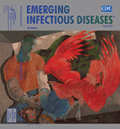
And the Raven, Never Flitting, Still Is Sitting, Still Is Sitting [PDF - 141 KB - 2 pages]
| EID | Potter P. And the Raven, Never Flitting, Still Is Sitting, Still Is Sitting. Emerg Infect Dis. 2010;16(10):1655-1656. https://doi.org/10.3201/eid1610.ac1610 |
|---|---|
| AMA | Potter P. And the Raven, Never Flitting, Still Is Sitting, Still Is Sitting. Emerging Infectious Diseases. 2010;16(10):1655-1656. doi:10.3201/eid1610.ac1610. |
| APA | Potter, P. (2010). And the Raven, Never Flitting, Still Is Sitting, Still Is Sitting. Emerging Infectious Diseases, 16(10), 1655-1656. https://doi.org/10.3201/eid1610.ac1610. |


