Volume 16, Number 12—December 2010
Dispatch
Emergence of African Swine Fever Virus, Northwestern Iran
Figure
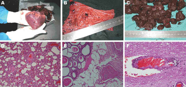
Figure. Acute form of African swine fever in wild boars. A) Petechial and larger ecchymotic hemorrhages beneath the epicardium. B) Severe hyperemia and petechial and larger ecchymotic hemorrhages in mucosa of urinary bladder. These hemorrhages are common in acute infectious fever and hemorrhagic diathesis. C) Blood-tinged colon contents with fecal balls covered by thick, blood-stained mucus. D) Congestion and fibrinous thromboses in pulmonary vessels and thickening of alveoli (hematoxylin and eosin stain; original magnification ×100). E) Fibrinous thrombus in a venule within interlobular adipose tissue of the thyroid gland (hematoxylin and eosin stain; original magnification ×200). F) Blood vessel congestion, perivascular hemorrhage, lymphocytic perivascular cuffing, and infiltration with degenerating lymphocytes (hematoxylin and eosin stain; original magnification ×200).