Volume 16, Number 12—December 2010
Letter
Brucellosis Reactivation after 28 Years
Figure
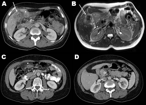
Figure. A) Contrast-enhanced computerized tomography (CT) scan showing a calcified gallbladder wall (arrow), a surrounding, calcified mass located peripherally in the liver, and an abscess in the adjacent fat tissue (arrowhead). B) T2-weighted axial magnetic resonance imaging shows multiple gallstones and a thickened gallbladder wall (arrow), inflammation and edema of the adjacent liver, fat tissue, and proximal duodenum. C) Eight weeks after cholecystectomy, contrast-enhanced CT shows a residual abscess in the adjacent fat tissue (arrowhead). D) Contrast-enhanced CT 5 months after cholecystectomy shows only minimal changes in the gallbladder bed and surrounding tissues, and no residual abscess.
1These authors contributed equally to this article.