Volume 16, Number 2—February 2010
Letter
Bronchial Casts and Pandemic (H1N1) 2009 Virus Infection
Maki Hasegawa, Yasuji Inamo , Tatsuo Fuchigami, Koji Hashimoto, Miyuki Morozumi, Kimiko Ubukata, Haruo Watanabe, and Takashi Takahashi
Figure
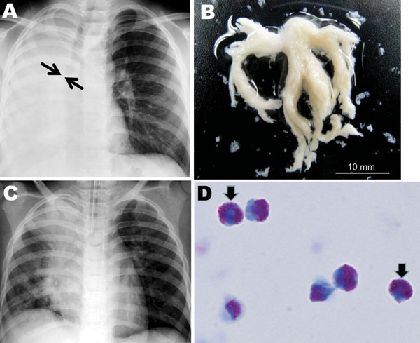
Figure. A) Chest radiograph obtained at hospital admission from a child infected with influenza subtype H1N1 virus. The image shows atelectasis of the right lung and hyperinflation of the left lung; arrows indicate obstruction of the right main bronchus. B) Macroscopic bronchial casts extracted by intratracheal suction. C) Chest radiograph obtained on hospital day 2, indicating partial resolution of atelectasis of the right lower lobe. D) Light micrograph of casts, characterized by predominant eosinophil infiltration (>90% of cells) (May-Giemsa stain, original magnification ×1,000). Arrows indicate typical eosinophil granules.
Page created: December 13, 2010
Page updated: December 13, 2010
Page reviewed: December 13, 2010
The conclusions, findings, and opinions expressed by authors contributing to this journal do not necessarily reflect the official position of the U.S. Department of Health and Human Services, the Public Health Service, the Centers for Disease Control and Prevention, or the authors' affiliated institutions. Use of trade names is for identification only and does not imply endorsement by any of the groups named above.