Volume 16, Number 7—July 2010
Dispatch
Accumulation of L-type Bovine Prions in Peripheral Nerve Tissues
Figure 1
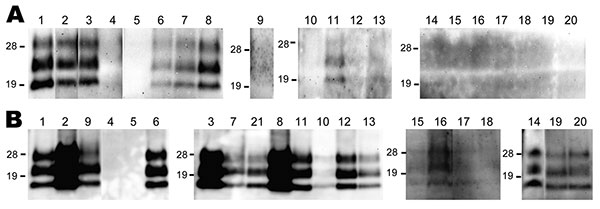
Figure 1. Western blot analysis of a protease-resistant form (PrPres) of a normal cellular prion protein in nerve tissue samples obtained from cattle 10 (A) and 16 (B) months postinoculation (cattle identification codes 8515 and 1061, respectively). The nerve tissues tested are shown above the lanes: 1, trigeminal ganglia; 2, pituitary gland; 3, anterior cervical ganglion; 4, facial nerve; 5, hypoglossal nerve; 6, cranial mesenteric ganglia; 7, vagus nerve (cervical part); 8, stellate ganglia; 9, adrenal gland; 10, phrenic nerve; 11, vagus nerve (pectoral part); 12, vagosympathic trunk (pectoral part); 13, vagosympathetic trunk (lumbar part); 14, accessory nerve; 15, suprascapular nerve; 16, brachial nerve plexus; 17, median nerve; 18, radial nerve; 19, sciatic nerve; 20, tibial nerve, 21, middle cervical ganglion. The equivalent of 100 mg of tissue was loaded. Western blots were probed with monoclonal antibody T2 to detect PrPres. Molecular mass standards (kDa) are indicated on the left of each panel.