Volume 17, Number 11—November 2011
Dispatch
Seasonal Influenza A Virus in Feces of Hospitalized Adults
Figure 2
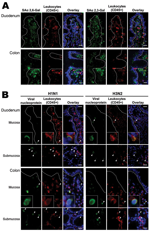
Figure 2. Intestinal distribution of influenza virus receptors and in vitro binding of inactivated seasonal influenza A (H1N1) and A (H3N2) viruses to human duodenal and colonic tissues. Images in the panels labeled Overlay show the green, red, and blue (nuclei counterstain) color channels in the same view. Dotted lines outline basal lining of intestinal epithelium. Arrowheads denote virus-bound cells. Scale bars = 20 µm. A) Double immunofluorescence staining showing that human-like influenza virus receptor sialic acid (SA) α 2,6-Gal; green) was not found on epithelial surface of small and large intestines but in lamina propria cells. Avian-like influenza virus receptor (SAα 2,3-Gal; green) was found on colonic epithelial surface and in lamina propria cells. Part of receptor-positive cells coexpressed CD45 (leukocyte common antigen; red), representing leukocytes. B) In vitro virus binding showing that neither seasonal influenza A (H1N1) nor A (H3N2) viruses bind to epithelial surface of small and large intestines but only to a subset of intestinal CD45+ leukocytes interspersed in the lamina propria and submucosa.
1These authors contributed equally to this article.