Volume 17, Number 12—December 2011
Research
Enterovirus Co-infections and Onychomadesis after Hand, Foot, and Mouth Disease, Spain, 2008
Maria A. Bracho , Fernando González-Candelas, Ana Valero, Juan Córdoba, and Antonio Salazar
Figure 3
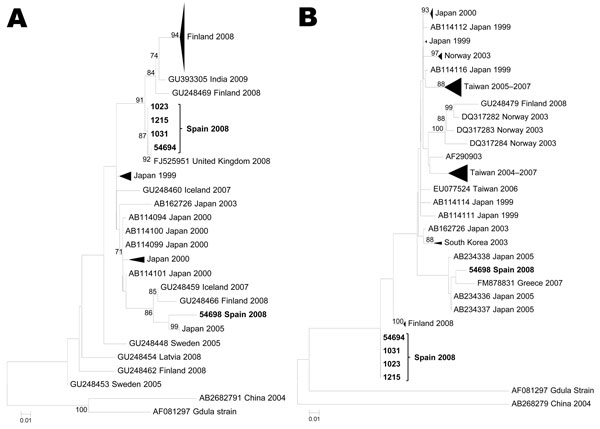
Figure 3. Maximum likelihood phylogenetic reconstructions for coxsackievirus A6 based on partial viral protein 1 sequences. A) 5′ partial coding region (81 strains, 293 nt). B) 3′ partial coding region (68 sequences, 377 nt). Bootstrap values >75% are shown. Scale bars indicate number of substitutions per nucleotide position. Multiple strains from the same country sharing the same node were collapsed and shown as triangles with shape proportional to branch distances and number of sequences.
Page created: November 30, 2011
Page updated: November 30, 2011
Page reviewed: November 30, 2011
The conclusions, findings, and opinions expressed by authors contributing to this journal do not necessarily reflect the official position of the U.S. Department of Health and Human Services, the Public Health Service, the Centers for Disease Control and Prevention, or the authors' affiliated institutions. Use of trade names is for identification only and does not imply endorsement by any of the groups named above.