Volume 17, Number 12—December 2011
Dispatch
Characterization of African Swine Fever Virus Caucasus Isolate in European Wild Boars
Claudia Gabriel, Sandra Blome , Alexander Malogolovkin, Stanislav Parilov, Denis Kolbasov, Jens P. Teifke, and Martin Beer
Figure 2
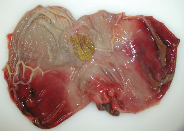
Figure 2. View of the mucosal surface of the dissected stomach showing representative gross lesions after oral inoculation of a wild boar with 106 median tissue culture infectious dose of an African swine fever virus isolate from Armenia (experiment at the Friedrich-Loeffler-Institut). The image illustrates acute gastritis; note diffuse mucosal hemorrhages affecting a large part of the mucosa. The animal died on day 7 postinfection.
Page created: November 30, 2011
Page updated: November 30, 2011
Page reviewed: November 30, 2011
The conclusions, findings, and opinions expressed by authors contributing to this journal do not necessarily reflect the official position of the U.S. Department of Health and Human Services, the Public Health Service, the Centers for Disease Control and Prevention, or the authors' affiliated institutions. Use of trade names is for identification only and does not imply endorsement by any of the groups named above.