Blastomycosis in Man after Kinkajou Bite
Julie R. Harris

, David D. Blaney, Mark D. Lindsley, Sherif R. Zaki, Christopher D. Paddock, Clifton P. Drew, April J. Johnson, Douglas Landau, Joel Vanderbush, and Robert Baker
Author affiliations: Author affiliations: Centers for Disease Control and Prevention, Atlanta, Georgia, USA (J.R. Harris, D.D. Blaney, M.D. Lindsley, S.A. Zaki, C.D. Paddock, C.P. Drew); Purdue University School of Veterinary Medicine, West Lafayette, Indiana, USA (A.J. Johnson); Indiana State Department of Health Laboratories, Indianapolis, Indiana, USA (D. Landau); Animalia, Inc., Indianapolis (J. Vanderbush); Community Health Network, Indianapolis (R. Baker)
Main Article
Figure 2
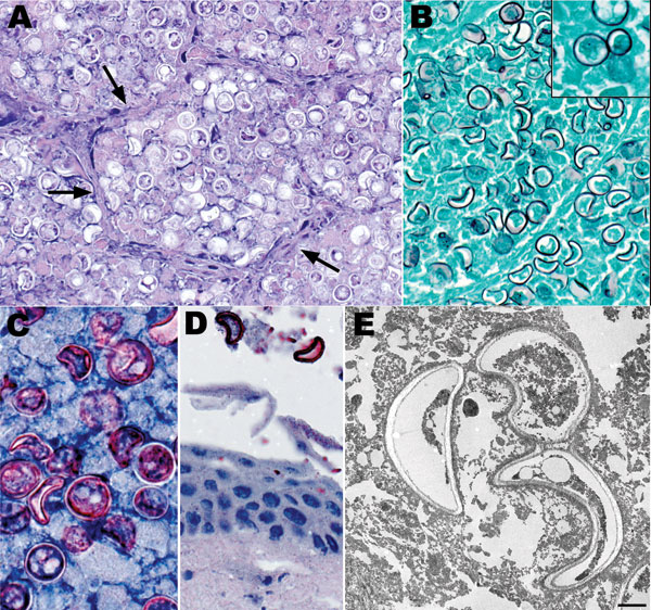
Figure 2. Histopathologic and electron microscopic appearance of Blastomyces dermatitidis in kinkajou (Potos flavus) tissues. A) Lung showing B. dermatitidis yeast forms filling alveolar spaces. Alveolar septa are indicated by arrows. B) Lung showing yeast forms of B. dermatitidis. Inset shows broad-based budding of a yeast form, a major diagnostic feature. C) Lung showing B. dermatitidis yeast. D) Oral mucosa showing 2 yeast forms of B. dermatitidis closely associated with the mucosal surface. E) Transmission electron micrograph showing 3 yeast forms of B. dermatitidis in lung tissue. Note the thick cell walls and crescent shapes of the yeast (scale bar = 2 µm). Hematoxylin and eosin stain (A), Grocott methenamine silver stain (B and inset), and immunoalkaline phosphatase with antibody against B. dermatitidis and naphthol fast-red with hematoxylin counterstain (C, D). Original magnifications ×400 (A, B, D) and ×630 (Inset, C).
Main Article
Page created: July 13, 2011
Page updated: July 13, 2011
Page reviewed: July 13, 2011
The conclusions, findings, and opinions expressed by authors contributing to this journal do not necessarily reflect the official position of the U.S. Department of Health and Human Services, the Public Health Service, the Centers for Disease Control and Prevention, or the authors' affiliated institutions. Use of trade names is for identification only and does not imply endorsement by any of the groups named above.