Volume 18, Number 10—October 2012
Dispatch
Powassan Virus Encephalitis, Minnesota, USA
Figure 1
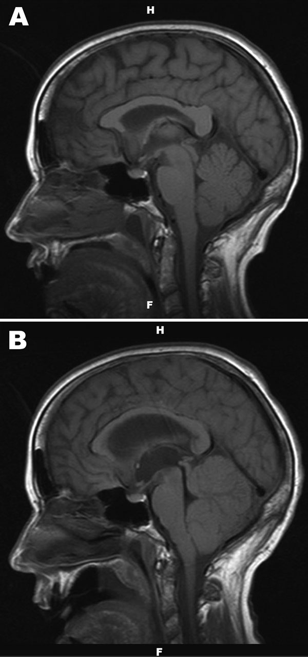
Figure 1. . . A) Noncontrast, sagittal T1-weighted magnetic resonance image of the brain of a 67-year-old woman with suspected Powassan virus encephalitis, obtained 4 days after admission. Image is notable for nonspecific signal changes within the thalami, midbrain, cerebellar vermis, and both cerebellar hemispheres. B) Noncontrast, sagittal T1-weighted magnetic resonance image of the brain obtained 8 days after patient’s admission. Changes include marked interval progression of signal abnormality involving the cerebellum, thalamus, and midbrain. Mass effect within the posterior fossa and crowding of structures at the foramen magnum are also evident. Marked dilatation of the lateral and third ventricles with acute hydrocephalus is apparent.