Research
Constant Transmission Properties of Variant Creutzfeldt-Jakob Disease in 5 Countries [PDF - 274 KB - 6 pages]
Variant Creutzfeldt-Jakob disease (vCJD) has been reported in 12 countries. We hypothesized that a common strain of agent is responsible for all vCJD cases, regardless of geographic origin. To test this hypothesis, we inoculated strain-typing panels of wild-type mice with brain material from human vCJD case-patients from France, the Netherlands, Italy, and the United States. Mice were assessed for clinical disease, neuropathologic changes, and glycoform profile; results were compared with those for 2 reference vCJD cases from the United Kingdom. Transmission to mice occurred from each sample tested, and data were similar between non-UK and UK cases, with the exception of the ranking of mean clinical incubation times of mouse lines. These findings support the hypothesis that a single strain of infectious agent is responsible for all vCJD infections. However, differences in incubation times require further subpassage in mice to establish any true differences in strain properties between cases.
| EID | Diack AB, Ritchie D, Bishop M, Pinion V, Brandel J, Haik S, et al. Constant Transmission Properties of Variant Creutzfeldt-Jakob Disease in 5 Countries. Emerg Infect Dis. 2012;18(10):1574-1579. https://doi.org/10.3201/eid1810.120792 |
|---|---|
| AMA | Diack AB, Ritchie D, Bishop M, et al. Constant Transmission Properties of Variant Creutzfeldt-Jakob Disease in 5 Countries. Emerging Infectious Diseases. 2012;18(10):1574-1579. doi:10.3201/eid1810.120792. |
| APA | Diack, A. B., Ritchie, D., Bishop, M., Pinion, V., Brandel, J., Haik, S....Manson, J. C. (2012). Constant Transmission Properties of Variant Creutzfeldt-Jakob Disease in 5 Countries. Emerging Infectious Diseases, 18(10), 1574-1579. https://doi.org/10.3201/eid1810.120792. |
WU and KI Polyomaviruses in Respiratory Samples from Allogeneic Hematopoietic Cell Transplant Recipients [PDF - 295 KB - 9 pages]
Data are limited regarding 2 new human polyomaviruses, KI polyomavirus (KIPyV) and WU polyomavirus (WUPyV), in immunocompromised patients. We used real-time PCR to test for these and 12 respiratory viruses in 2,732 nasal wash samples collected during the first year after allogeneic hematopoietic cell transplantation from 222 patients. Specimens were collected weekly until day 100; then at least every 3 months. One year after hematopoietic cell transplantation, the cumulative incidence estimate was 26% for KIPyV and 8% for WUPyV. Age <20 years predicted detection of KIPyV (hazard ratio [HR] 4.6) and WUPyV (HR 4.4), and detection of a respiratory virus in the previous 2 weeks predicted KIPyV detection (HR 3.4). Sputum production and wheezing were associated with detection of KIPyV in the past week and WUPyV in the past month. There were no associations with polyomavirus detection and acute graft versus host disease, cytomegalovirus reactivation, neutropenia, lymphopenia, hospitalization, or death.
| EID | Kuypers J, Campbell AP, Guthrie KA, Wright NL, Englund JA, Corey L, et al. WU and KI Polyomaviruses in Respiratory Samples from Allogeneic Hematopoietic Cell Transplant Recipients. Emerg Infect Dis. 2012;18(10):1580-1588. https://doi.org/10.3201/eid1810.120477 |
|---|---|
| AMA | Kuypers J, Campbell AP, Guthrie KA, et al. WU and KI Polyomaviruses in Respiratory Samples from Allogeneic Hematopoietic Cell Transplant Recipients. Emerging Infectious Diseases. 2012;18(10):1580-1588. doi:10.3201/eid1810.120477. |
| APA | Kuypers, J., Campbell, A. P., Guthrie, K. A., Wright, N. L., Englund, J. A., Corey, L....Boeckh, M. (2012). WU and KI Polyomaviruses in Respiratory Samples from Allogeneic Hematopoietic Cell Transplant Recipients. Emerging Infectious Diseases, 18(10), 1580-1588. https://doi.org/10.3201/eid1810.120477. |
Wild Birds and Urban Ecology of Ticks and Tick-borne Pathogens, Chicago, Illinois, USA, 2005–2010 [PDF - 239 KB - 7 pages]
Bird-facilitated introduction of ticks and associated pathogens is postulated to promote invasion of tick-borne zoonotic diseases into urban areas. Results of a longitudinal study conducted in suburban Chicago, Illinois, USA, during 2005–2010 show that 1.6% of 6,180 wild birds captured in mist nets harbored ticks. Tick species in order of abundance were Haemaphysalis leporispalustris, Ixodes dentatus, and I. scapularis, but 2 neotropical tick species of the genus Amblyomma were sampled during the spring migration. I. scapularis ticks were absent at the beginning of the study but constituted the majority of ticks by study end and were found predominantly on birds captured in areas designated as urban green spaces. Of 120 ticks, 5 were infected with Borrelia burgdorferi, spanning 3 ribotypes, but none were infected with Anaplasma phagocytophilum. Results allow inferences about propagule pressure for introduction of tick-borne diseases and emphasize the large sample sizes required to estimate this pressure.
| EID | Hamer SA, Goldberg TL, Kitron UD, Brawn JD, Anderson TK, Loss SR, et al. Wild Birds and Urban Ecology of Ticks and Tick-borne Pathogens, Chicago, Illinois, USA, 2005–2010. Emerg Infect Dis. 2012;18(10):1589-1595. https://doi.org/10.3201/eid1810.120511 |
|---|---|
| AMA | Hamer SA, Goldberg TL, Kitron UD, et al. Wild Birds and Urban Ecology of Ticks and Tick-borne Pathogens, Chicago, Illinois, USA, 2005–2010. Emerging Infectious Diseases. 2012;18(10):1589-1595. doi:10.3201/eid1810.120511. |
| APA | Hamer, S. A., Goldberg, T. L., Kitron, U. D., Brawn, J. D., Anderson, T. K., Loss, S. R....Hamer, G. L. (2012). Wild Birds and Urban Ecology of Ticks and Tick-borne Pathogens, Chicago, Illinois, USA, 2005–2010. Emerging Infectious Diseases, 18(10), 1589-1595. https://doi.org/10.3201/eid1810.120511. |
Spread of Influenza Virus A (H5N1) Clade 2.3.2.1 to Bulgaria in Common Buzzards [PDF - 287 KB - 7 pages]
On March 15, 2010, a highly pathogenic avian influenza virus was isolated from the carcass of a common buzzard (Buteo buteo) in Bulgaria. Phylogenetic analyses of the virus showed a close genetic relationship with influenza virus A (H5N1) clade 2.3.2.1 viruses isolated from wild birds in the Tyva Republic and Mongolia during 2009–2010. Designated A/common buzzard/Bulgaria/38WB/2010, this strain was highly pathogenic in chickens but had low pathogenicity in mice and ferrets and no molecular markers of increased pathogenicity in mammals. The establishment of clade 2.3.2.1 highly pathogenic avian influenza viruses of the H5N1 subtype in wild birds in Europe would increase the likelihood of health threats to humans and poultry in the region.
| EID | Marinova-Petkova A, Georgiev G, Seiler P, Darnell D, Franks J, Krauss S, et al. Spread of Influenza Virus A (H5N1) Clade 2.3.2.1 to Bulgaria in Common Buzzards. Emerg Infect Dis. 2012;18(10):1596-1602. https://doi.org/10.3201/eid1810.120357 |
|---|---|
| AMA | Marinova-Petkova A, Georgiev G, Seiler P, et al. Spread of Influenza Virus A (H5N1) Clade 2.3.2.1 to Bulgaria in Common Buzzards. Emerging Infectious Diseases. 2012;18(10):1596-1602. doi:10.3201/eid1810.120357. |
| APA | Marinova-Petkova, A., Georgiev, G., Seiler, P., Darnell, D., Franks, J., Krauss, S....Webster, R. G. (2012). Spread of Influenza Virus A (H5N1) Clade 2.3.2.1 to Bulgaria in Common Buzzards. Emerging Infectious Diseases, 18(10), 1596-1602. https://doi.org/10.3201/eid1810.120357. |
Dengue Outbreaks in High-Income Area, Kaohsiung City, Taiwan, 2003–2009 [PDF - 346 KB - 9 pages]
Kaohsiung City, a modern metropolis of 1.5 million persons, has been the focus of dengue virus activity in Taiwan for several decades. The aim of this study was to provide a temporal and spatial description of dengue virus epidemiology in Kaohsiung City by using data for all laboratory-confirmed dengue cases during 2003–2009. We investigated age- and sex-dependent incidence rates and the spatiotemporal patterns of all cases confirmed through passive or active surveillance. Elderly persons were at particularly high risk for dengue virus–related sickness and death. Of all confirmed cases, ≈75% were detected through passive surveillance activities; case-patients detected through active surveillance included immediate family members, neighbors, and colleagues of confirmed case-patients. Changing patterns of case clustering could be due to the effect of unmeasured environmental and demographic factors.
| EID | Lin C, Schiøler KL, Jepsen MR, Ho C, Li S, Konradsen F. Dengue Outbreaks in High-Income Area, Kaohsiung City, Taiwan, 2003–2009. Emerg Infect Dis. 2012;18(10):1603-1611. https://doi.org/10.3201/eid1810.111929 |
|---|---|
| AMA | Lin C, Schiøler KL, Jepsen MR, et al. Dengue Outbreaks in High-Income Area, Kaohsiung City, Taiwan, 2003–2009. Emerging Infectious Diseases. 2012;18(10):1603-1611. doi:10.3201/eid1810.111929. |
| APA | Lin, C., Schiøler, K. L., Jepsen, M. R., Ho, C., Li, S., & Konradsen, F. (2012). Dengue Outbreaks in High-Income Area, Kaohsiung City, Taiwan, 2003–2009. Emerging Infectious Diseases, 18(10), 1603-1611. https://doi.org/10.3201/eid1810.111929. |
Nontuberculous Mycobacteria in Household Plumbing as Possible Cause of Chronic Rhinosinusitis [PDF - 194 KB - 6 pages]
Symptoms of chronic rhinosinusitis (CRS) often persist despite treatment. Because nontuberculous mycobacteria (NTM) are resistant to commonly used antimicrobial drugs and are found in drinking water that patients may use for sinus irrigation, we investigated whether some CRS patients were infected with NTM in New York, New York, USA, during 2001–2011. Two approaches were chosen: 1) records of NTM-infected CRS patients were reviewed to identify common features of infection and Mycobacterium species; 2) samples from plumbing in households of 8 NTM-infected patients were cultured for NTM presence. In 3 households sampled, M. avium sharing rep-PCR and pulsed field gel electrophoresis fingerprints identified M. avium isolates clonally related to the patients’ isolates. We conclude that patients with treatment-resistant CRS may be infected with NTM and should have cultures performed for NTM so appropriate therapy can be instituted. In addition, the results suggest that CRS patients can be infected by NTM in their household plumbing.
| EID | Tichenor WS, Thurlow J, McNulty S, Brown-Elliott BA, Wallace RJ, Falkinham JO. Nontuberculous Mycobacteria in Household Plumbing as Possible Cause of Chronic Rhinosinusitis. Emerg Infect Dis. 2012;18(10):1612-1617. https://doi.org/10.3201/eid1810.120164 |
|---|---|
| AMA | Tichenor WS, Thurlow J, McNulty S, et al. Nontuberculous Mycobacteria in Household Plumbing as Possible Cause of Chronic Rhinosinusitis. Emerging Infectious Diseases. 2012;18(10):1612-1617. doi:10.3201/eid1810.120164. |
| APA | Tichenor, W. S., Thurlow, J., McNulty, S., Brown-Elliott, B. A., Wallace, R. J., & Falkinham, J. O. (2012). Nontuberculous Mycobacteria in Household Plumbing as Possible Cause of Chronic Rhinosinusitis. Emerging Infectious Diseases, 18(10), 1612-1617. https://doi.org/10.3201/eid1810.120164. |
Autochthonous and Dormant Cryptococcus gattii Infections in Europe [PDF - 217 KB - 7 pages]
Until recently, Cryptococcus gattii infections occurred mainly in tropical and subtropical climate zones. However, during the past decade, C. gattii infections in humans and animals in Europe have increased. To determine whether the infections in Europe were acquired from an autochthonous source or associated with travel, we used multilocus sequence typing to compare 100 isolates from Europe (57 from 40 human patients, 22 from the environment, and 21 from animals) with 191 isolates from around the world. Of the 57 human patient isolates, 47 (83%) were obtained since 1995. Among the 40 patients, 24 (60%) probably acquired the C. gattii infection outside Europe; the remaining 16 (40%) probably acquired the infection within Europe. Human patient isolates from Mediterranean Europe clustered into a distinct genotype with animal and environmental isolates. These results indicate that reactivation of dormant C. gattii infections can occur many years after the infectious agent was acquired elsewhere.
| EID | Hagen F, Colom M, Swinne D, Tintelnot K, Iatta R, Montagna M, et al. Autochthonous and Dormant Cryptococcus gattii Infections in Europe. Emerg Infect Dis. 2012;18(10):1618-1624. https://doi.org/10.3201/eid1810.120068 |
|---|---|
| AMA | Hagen F, Colom M, Swinne D, et al. Autochthonous and Dormant Cryptococcus gattii Infections in Europe. Emerging Infectious Diseases. 2012;18(10):1618-1624. doi:10.3201/eid1810.120068. |
| APA | Hagen, F., Colom, M., Swinne, D., Tintelnot, K., Iatta, R., Montagna, M....Boekhout, T. (2012). Autochthonous and Dormant Cryptococcus gattii Infections in Europe. Emerging Infectious Diseases, 18(10), 1618-1624. https://doi.org/10.3201/eid1810.120068. |
Noroviruses are the leading cause of foodborne illness in the United States. To better guide interventions, we analyzed 2,922 foodborne disease outbreaks for which norovirus was the suspected or confirmed cause, which had been reported to the Foodborne Disease Outbreak Surveillance System of the Centers for Disease Control and Prevention during 2001–2008. On average, 365 foodborne norovirus outbreaks were reported annually, resulting in an estimated 10,324 illnesses, 1,247 health care provider visits, 156 hospitalizations, and 1 death. In 364 outbreaks attributed to a single commodity, leafy vegetables (33%), fruits/nuts (16%), and mollusks (13%) were implicated most commonly. Infected food handlers were the source of 53% of outbreaks and may have contributed to 82% of outbreaks. Most foods were likely contaminated during preparation and service, except for mollusks, and occasionally, produce was contaminated during production and processing. Interventions to reduce the frequency of foodborne norovirus outbreaks should focus on food workers and production of produce and shellfish.
| EID | Hall AJ, Eisenbart VG, Etingüe A, Gould L, Lopman BA, Parashar UD. Epidemiology of Foodborne Norovirus Outbreaks, United States, 2001–2008. Emerg Infect Dis. 2012;18(10):1566-1573. https://doi.org/10.3201/eid1810.120833 |
|---|---|
| AMA | Hall AJ, Eisenbart VG, Etingüe A, et al. Epidemiology of Foodborne Norovirus Outbreaks, United States, 2001–2008. Emerging Infectious Diseases. 2012;18(10):1566-1573. doi:10.3201/eid1810.120833. |
| APA | Hall, A. J., Eisenbart, V. G., Etingüe, A., Gould, L., Lopman, B. A., & Parashar, U. D. (2012). Epidemiology of Foodborne Norovirus Outbreaks, United States, 2001–2008. Emerging Infectious Diseases, 18(10), 1566-1573. https://doi.org/10.3201/eid1810.120833. |
Methicillin-resistant Staphylococcus aureus (MRSA) is a human pathogen that has diverse molecular heterogeneity. Most MRSA strains in the United States are pulsed-field gel electrophoresis USA100 sequence type (ST) 5 and USA300 ST8. Infections with MRSA ST239-III are common and found during health care–associated outbreaks. However, this strain has been rarely reported in the United States. As part of a study supported by the Prevention Epicenter Program of the Centers for Disease Control and Prevention (Atlanta, GA, USA), which evaluated transmission of MRSA among hospitals in Ohio, molecular typing identified 78 (6%) of 1,286 patients with MRSA ST239-III infections. Ninety-five percent (74/78) of these infections were health care associated, and 65% (51/78) of patients had histories of invasive device use. The crude case-fatality rate was 22% (17/78). Identification of these strains, which belong to a virulent clonal group, emphasizes the need for molecular surveillance.
| EID | Wang S, Khan Y, Hines L, Mediavilla JR, Zhang L, Chen L, et al. Methicillin-Resistant Staphylococcus aureus Sequence Type 239-III, Ohio, USA, 2007–2009. Emerg Infect Dis. 2012;18(10):1557-1565. https://doi.org/10.3201/eid1810.120468 |
|---|---|
| AMA | Wang S, Khan Y, Hines L, et al. Methicillin-Resistant Staphylococcus aureus Sequence Type 239-III, Ohio, USA, 2007–2009. Emerging Infectious Diseases. 2012;18(10):1557-1565. doi:10.3201/eid1810.120468. |
| APA | Wang, S., Khan, Y., Hines, L., Mediavilla, J. R., Zhang, L., Chen, L....Stevenson, K. B. (2012). Methicillin-Resistant Staphylococcus aureus Sequence Type 239-III, Ohio, USA, 2007–2009. Emerging Infectious Diseases, 18(10), 1557-1565. https://doi.org/10.3201/eid1810.120468. |
Dispatches
Echinococcus multilocularis in Urban Coyotes, Alberta, Canada [PDF - 355 KB - 4 pages]
Echinococcus multilocularis is a zoonotic parasite in wild canids. We determined its frequency in urban coyotes (Canis latrans) in Alberta, Canada. We detected E. multilocularis in 23 of 91 coyotes in this region. This parasite is a public health concern throughout the Northern Hemisphere, partly because of increased urbanization of wild canids.
| EID | Catalano S, Lejeune M, Liccioli S, Verocai GG, Gesy KM, Jenkins EJ, et al. Echinococcus multilocularis in Urban Coyotes, Alberta, Canada. Emerg Infect Dis. 2012;18(10):1625-1628. https://doi.org/10.3201/eid1810.120119 |
|---|---|
| AMA | Catalano S, Lejeune M, Liccioli S, et al. Echinococcus multilocularis in Urban Coyotes, Alberta, Canada. Emerging Infectious Diseases. 2012;18(10):1625-1628. doi:10.3201/eid1810.120119. |
| APA | Catalano, S., Lejeune, M., Liccioli, S., Verocai, G. G., Gesy, K. M., Jenkins, E. J....Massolo, A. (2012). Echinococcus multilocularis in Urban Coyotes, Alberta, Canada. Emerging Infectious Diseases, 18(10), 1625-1628. https://doi.org/10.3201/eid1810.120119. |
Orthobunyavirus Antibodies in Humans, Yucatan Peninsula, Mexico [PDF - 308 KB - 4 pages]
We performed a serologic investigation to determine whether orthobunyaviruses commonly infect humans in the Yucatan Peninsula of Mexico. Orthobunyavirus-specific antibodies were detected by plaque reduction neutralization test in 146 (18%) of 823 persons tested. Further studies are needed to determine health risks for humans from this potentially deadly group of viruses.
| EID | Blitvich BJ, Saiyasombat R, Talavera-Aguilar LG, Garcia-Rejon JE, Farfan-Ale JA, Machain-Williams C, et al. Orthobunyavirus Antibodies in Humans, Yucatan Peninsula, Mexico. Emerg Infect Dis. 2012;18(10):1629-1632. https://doi.org/10.3201/eid1810.120492 |
|---|---|
| AMA | Blitvich BJ, Saiyasombat R, Talavera-Aguilar LG, et al. Orthobunyavirus Antibodies in Humans, Yucatan Peninsula, Mexico. Emerging Infectious Diseases. 2012;18(10):1629-1632. doi:10.3201/eid1810.120492. |
| APA | Blitvich, B. J., Saiyasombat, R., Talavera-Aguilar, L. G., Garcia-Rejon, J. E., Farfan-Ale, J. A., Machain-Williams, C....Loroño-Pino, M. A. (2012). Orthobunyavirus Antibodies in Humans, Yucatan Peninsula, Mexico. Emerging Infectious Diseases, 18(10), 1629-1632. https://doi.org/10.3201/eid1810.120492. |
Tetanus as Cause of Mass Die-off of Captive Japanese Macaques, Japan, 2008 [PDF - 204 KB - 3 pages]
In 2008 in Japan, 15/60 captive Japanese macaques died. Clostridium tetani was isolated from 1 monkey, and 11 had tetanus-specific symptoms. We conclude the outbreak resulted from severe environmental C. tetani contamination. Similar outbreaks could be prevented by vaccinating all monkeys, disinfecting housing areas/play equipment, replacing highly C. tetani–contaminated soil, and conducting epidemiologic surveys.
| EID | Nakano T, Nakamura S, Yamamoto A, Takahashi M, Une Y. Tetanus as Cause of Mass Die-off of Captive Japanese Macaques, Japan, 2008. Emerg Infect Dis. 2012;18(10):1633-1635. https://doi.org/10.3201/eid1810.120503 |
|---|---|
| AMA | Nakano T, Nakamura S, Yamamoto A, et al. Tetanus as Cause of Mass Die-off of Captive Japanese Macaques, Japan, 2008. Emerging Infectious Diseases. 2012;18(10):1633-1635. doi:10.3201/eid1810.120503. |
| APA | Nakano, T., Nakamura, S., Yamamoto, A., Takahashi, M., & Une, Y. (2012). Tetanus as Cause of Mass Die-off of Captive Japanese Macaques, Japan, 2008. Emerging Infectious Diseases, 18(10), 1633-1635. https://doi.org/10.3201/eid1810.120503. |
Human Infection with Candidatus Neoehrlichia mikurensis, China [PDF - 344 KB - 4 pages]
To identify Candidatus Neoehrlichia mikurensis infection in northeastern China, we tested blood samples from 622 febrile patients. We identified in 7 infected patients and natural foci for this bacterium. Field surveys showed that 1.6% of ticks and 3.8% of rodents collected from residences of patients were also infected.
| EID | Li H, Jiang J, Liu W, Zheng Y, Huo Q, Tang K, et al. Human Infection with Candidatus Neoehrlichia mikurensis, China. Emerg Infect Dis. 2012;18(10):1636-1639. https://doi.org/10.3201/eid1810.120594 |
|---|---|
| AMA | Li H, Jiang J, Liu W, et al. Human Infection with Candidatus Neoehrlichia mikurensis, China. Emerging Infectious Diseases. 2012;18(10):1636-1639. doi:10.3201/eid1810.120594. |
| APA | Li, H., Jiang, J., Liu, W., Zheng, Y., Huo, Q., Tang, K....Cao, W. (2012). Human Infection with Candidatus Neoehrlichia mikurensis, China. Emerging Infectious Diseases, 18(10), 1636-1639. https://doi.org/10.3201/eid1810.120594. |
Anthroponotic Enteric Parasites in Monkeys in Public Park, China [PDF - 357 KB - 4 pages]
Cryptosporidium spp., Giardia duodenalis, and Enterocytozoon bieneusi were detected in 45, 35, and 116 of 411 free-range rhesus monkeys, respectively, in a popular public park in the People’s Republic of China. Most genotypes and subtypes detected were anthroponotic, indicating these animals might be reservoirs for human cryptosporidiosis, giardiasis, and microsporidiosis.
| EID | Ye J, Xiao L, Ma J, Guo M, Liu L, Feng Y. Anthroponotic Enteric Parasites in Monkeys in Public Park, China. Emerg Infect Dis. 2012;18(10):1640-1643. https://doi.org/10.3201/eid1810.120653 |
|---|---|
| AMA | Ye J, Xiao L, Ma J, et al. Anthroponotic Enteric Parasites in Monkeys in Public Park, China. Emerging Infectious Diseases. 2012;18(10):1640-1643. doi:10.3201/eid1810.120653. |
| APA | Ye, J., Xiao, L., Ma, J., Guo, M., Liu, L., & Feng, Y. (2012). Anthroponotic Enteric Parasites in Monkeys in Public Park, China. Emerging Infectious Diseases, 18(10), 1640-1643. https://doi.org/10.3201/eid1810.120653. |
Schmallenberg Virus as Possible Ancestor of Shamonda Virus [PDF - 222 KB - 3 pages]
Schmallenberg virus (SBV), an orthobunyavirus of the Simbu serogroup, recently emerged in Europe and has been suggested to be a Shamonda/Sathuperi virus reassortant. Results of full-genome and serologic investigations indicate that SBV belongs to the species Sathuperi virus and is a possible ancestor of the reassortant Shamonda virus.
| EID | Goller KV, Höper D, Schirrmeier H, Mettenleiter TC, Beer M. Schmallenberg Virus as Possible Ancestor of Shamonda Virus. Emerg Infect Dis. 2012;18(10):1644-1646. https://doi.org/10.3201/eid1810.120835 |
|---|---|
| AMA | Goller KV, Höper D, Schirrmeier H, et al. Schmallenberg Virus as Possible Ancestor of Shamonda Virus. Emerging Infectious Diseases. 2012;18(10):1644-1646. doi:10.3201/eid1810.120835. |
| APA | Goller, K. V., Höper, D., Schirrmeier, H., Mettenleiter, T. C., & Beer, M. (2012). Schmallenberg Virus as Possible Ancestor of Shamonda Virus. Emerging Infectious Diseases, 18(10), 1644-1646. https://doi.org/10.3201/eid1810.120835. |
Monkey Bites among US Military Members, Afghanistan, 2011 [PDF - 195 KB - 3 pages]
Bites from Macaca mulatta monkeys, native to Afghanistan, can cause serious infections. To determine risk for US military members in Afghanistan, we reviewed records for September–December 2011. Among 126 animal bites and exposures, 10 were monkey bites. Command emphasis is vital for preventing monkey bites; provider training and bite reporting promote postexposure treatment.
| EID | Mease LE, Baker KA. Monkey Bites among US Military Members, Afghanistan, 2011. Emerg Infect Dis. 2012;18(10):1647-1649. https://doi.org/10.3201/eid1810.120419 |
|---|---|
| AMA | Mease LE, Baker KA. Monkey Bites among US Military Members, Afghanistan, 2011. Emerging Infectious Diseases. 2012;18(10):1647-1649. doi:10.3201/eid1810.120419. |
| APA | Mease, L. E., & Baker, K. A. (2012). Monkey Bites among US Military Members, Afghanistan, 2011. Emerging Infectious Diseases, 18(10), 1647-1649. https://doi.org/10.3201/eid1810.120419. |
Hepatitis E Virus Seroprevalence among Adults, Germany [PDF - 285 KB - 4 pages]
We assessed hepatitis E virus (HEV) antibody seroprevalence in a sample of the adult population in Germany. Overall HEV IgG prevalence was 16.8% (95% CI 15.6%–17.9%) and increased with age, leveling off at >60 years of age. HEV is endemic in Germany, and the lifetime risk for exposure is high.
| EID | Faber MS, Wenzel JJ, Jilg W, Thamm M, Höhle M, Stark K. Hepatitis E Virus Seroprevalence among Adults, Germany. Emerg Infect Dis. 2012;18(10):1654-1657. https://doi.org/10.3201/eid1810.111756 |
|---|---|
| AMA | Faber MS, Wenzel JJ, Jilg W, et al. Hepatitis E Virus Seroprevalence among Adults, Germany. Emerging Infectious Diseases. 2012;18(10):1654-1657. doi:10.3201/eid1810.111756. |
| APA | Faber, M. S., Wenzel, J. J., Jilg, W., Thamm, M., Höhle, M., & Stark, K. (2012). Hepatitis E Virus Seroprevalence among Adults, Germany. Emerging Infectious Diseases, 18(10), 1654-1657. https://doi.org/10.3201/eid1810.111756. |
Scarlet Fever Epidemic, Hong Kong, 2011 [PDF - 298 KB - 4 pages]
More than 900 cases of scarlet fever were recorded in Hong Kong during January–July, 2011. Six cases were complicated by toxic shock syndrome, of which 2 were fatal. Pulsed-field gel electrophoresis patterns suggested a multiclonal epidemic; emm12 was the predominant circulating type. We recommend genetic testing of and antimicrobial resistance monitoring for this reportable disease.
| EID | Luk E, Lo J, Li A, Lau M, Cheung T, Wong A, et al. Scarlet Fever Epidemic, Hong Kong, 2011. Emerg Infect Dis. 2012;18(10):1658-1661. https://doi.org/10.3201/eid1810.111900 |
|---|---|
| AMA | Luk E, Lo J, Li A, et al. Scarlet Fever Epidemic, Hong Kong, 2011. Emerging Infectious Diseases. 2012;18(10):1658-1661. doi:10.3201/eid1810.111900. |
| APA | Luk, E., Lo, J., Li, A., Lau, M., Cheung, T., Wong, A....Tsang, T. (2012). Scarlet Fever Epidemic, Hong Kong, 2011. Emerging Infectious Diseases, 18(10), 1658-1661. https://doi.org/10.3201/eid1810.111900. |
Visceral Leishmaniasis in Rural Bihar, India [PDF - 243 KB - 3 pages]
To identify factors associated with incidence of visceral leishmaniasis (VL), we surveyed 13,416 households in Bihar State, India. VL was associated with socioeconomic status, type of housing, and belonging to the Musahar caste. Annual coverage of indoor residual insecticide spraying was 12%. Increasing such spraying can greatly contribute to VL control.
| EID | Hasker E, Singh S, Malaviya P, Picado A, Gidwani K, Singh R, et al. Visceral Leishmaniasis in Rural Bihar, India. Emerg Infect Dis. 2012;18(10):1662-1664. https://doi.org/10.3201/eid1810.111083 |
|---|---|
| AMA | Hasker E, Singh S, Malaviya P, et al. Visceral Leishmaniasis in Rural Bihar, India. Emerging Infectious Diseases. 2012;18(10):1662-1664. doi:10.3201/eid1810.111083. |
| APA | Hasker, E., Singh, S., Malaviya, P., Picado, A., Gidwani, K., Singh, R....Sundar, S. (2012). Visceral Leishmaniasis in Rural Bihar, India. Emerging Infectious Diseases, 18(10), 1662-1664. https://doi.org/10.3201/eid1810.111083. |
Influenza A(H1N1)pdm09 Virus in Pigs, Réunion Island [PDF - 342 KB - 4 pages]
During 2009, pandemic influenza A(H1N1)pdm09 virus affected humans on Réunion Island. Since then, the virus has sustained circulation among local swine herds, raising concerns about the potential for genetic evolution of the virus and possible retransmission back to humans of variants with increased virulence. Continuous surveillance of A(H1N1)pdm09 infection in pigs is recommended.
| EID | Cardinale E, Pascalis H, Temmam S, Hervé S, Saulnier A, Turpin M, et al. Influenza A(H1N1)pdm09 Virus in Pigs, Réunion Island. Emerg Infect Dis. 2012;18(10):1665-1668. https://doi.org/10.3201/eid1810.120398 |
|---|---|
| AMA | Cardinale E, Pascalis H, Temmam S, et al. Influenza A(H1N1)pdm09 Virus in Pigs, Réunion Island. Emerging Infectious Diseases. 2012;18(10):1665-1668. doi:10.3201/eid1810.120398. |
| APA | Cardinale, E., Pascalis, H., Temmam, S., Hervé, S., Saulnier, A., Turpin, M....Simon, G. (2012). Influenza A(H1N1)pdm09 Virus in Pigs, Réunion Island. Emerging Infectious Diseases, 18(10), 1665-1668. https://doi.org/10.3201/eid1810.120398. |
Powassan Virus Encephalitis, Minnesota, USA [PDF - 230 KB - 3 pages]
Powassan virus (POWV) is a rare tick-borne agent of encephalitis in North America. Historically, confirmed cases occurred mainly in the northeastern United States. Since 2008, confirmed cases in Minnesota and Wisconsin have increased. We report a fatal case of POWV encephalitis in Minnesota. POWV infection should be suspected in tick-exposed patients with viral encephalitis.
| EID | Birge J, Sonnesyn S. Powassan Virus Encephalitis, Minnesota, USA. Emerg Infect Dis. 2012;18(10):1669-1671. https://doi.org/10.3201/eid1810.120621 |
|---|---|
| AMA | Birge J, Sonnesyn S. Powassan Virus Encephalitis, Minnesota, USA. Emerging Infectious Diseases. 2012;18(10):1669-1671. doi:10.3201/eid1810.120621. |
| APA | Birge, J., & Sonnesyn, S. (2012). Powassan Virus Encephalitis, Minnesota, USA. Emerging Infectious Diseases, 18(10), 1669-1671. https://doi.org/10.3201/eid1810.120621. |
Influenza Virus Infection in Nonhuman Primates [PDF - 268 KB - 4 pages]
To determine whether nonhuman primates are infected with influenza viruses in nature, we conducted serologic and swab studies among macaques from several parts of the world. Our detection of influenza virus and antibodies to influenza virus raises questions about the role of nonhuman primates in the ecology of influenza.
| EID | Karlsson EA, Engel GA, Feeroz M, San S, Rompis A, Lee B, et al. Influenza Virus Infection in Nonhuman Primates. Emerg Infect Dis. 2012;18(10):1672-1675. https://doi.org/10.3201/eid1810.120214 |
|---|---|
| AMA | Karlsson EA, Engel GA, Feeroz M, et al. Influenza Virus Infection in Nonhuman Primates. Emerging Infectious Diseases. 2012;18(10):1672-1675. doi:10.3201/eid1810.120214. |
| APA | Karlsson, E. A., Engel, G. A., Feeroz, M., San, S., Rompis, A., Lee, B....Jones-Engel, L. (2012). Influenza Virus Infection in Nonhuman Primates. Emerging Infectious Diseases, 18(10), 1672-1675. https://doi.org/10.3201/eid1810.120214. |
Human Polyomaviruses in Children Undergoing Transplantation, United States, 2008–2010 [PDF - 206 KB - 4 pages]
Immunocompromised patients are at risk for disease caused by infection by some polyomaviruses. To define the prevalence of polyomaviruses in children undergoing transplantation, we collected samples from a longitudinal cohort and tested for the 9 known human polyomaviruses. All were detected; several were present in previously unreported specimen types.
| EID | Siebrasse EA, Bauer I, Holtz LR, Le B, Lassa-Claxton S, Canter C, et al. Human Polyomaviruses in Children Undergoing Transplantation, United States, 2008–2010. Emerg Infect Dis. 2012;18(10):1676-1679. https://doi.org/10.3201/eid1810.120359 |
|---|---|
| AMA | Siebrasse EA, Bauer I, Holtz LR, et al. Human Polyomaviruses in Children Undergoing Transplantation, United States, 2008–2010. Emerging Infectious Diseases. 2012;18(10):1676-1679. doi:10.3201/eid1810.120359. |
| APA | Siebrasse, E. A., Bauer, I., Holtz, L. R., Le, B., Lassa-Claxton, S., Canter, C....Wang, D. (2012). Human Polyomaviruses in Children Undergoing Transplantation, United States, 2008–2010. Emerging Infectious Diseases, 18(10), 1676-1679. https://doi.org/10.3201/eid1810.120359. |
Preventing Maritime Transfer of Toxigenic Vibrio cholerae [PDF - 215 KB - 3 pages]
Organisms, including Vibrio cholerae, can be transferred between harbors in the ballast water of ships. Zones in the Caribbean region where distance from shore and water depth meet International Maritime Organization guidelines for ballast water exchange are extremely limited. Use of ballast water treatment systems could mitigate the risk for organism transfer.
| EID | Cohen NJ, Slaten DD, Marano N, Tappero JW, Wellman M, Albert RJ, et al. Preventing Maritime Transfer of Toxigenic Vibrio cholerae. Emerg Infect Dis. 2012;18(10):1680-1682. https://doi.org/10.3201/eid1810.120676 |
|---|---|
| AMA | Cohen NJ, Slaten DD, Marano N, et al. Preventing Maritime Transfer of Toxigenic Vibrio cholerae. Emerging Infectious Diseases. 2012;18(10):1680-1682. doi:10.3201/eid1810.120676. |
| APA | Cohen, N. J., Slaten, D. D., Marano, N., Tappero, J. W., Wellman, M., Albert, R. J....Tauxe, R. V. (2012). Preventing Maritime Transfer of Toxigenic Vibrio cholerae. Emerging Infectious Diseases, 18(10), 1680-1682. https://doi.org/10.3201/eid1810.120676. |
Human Parvovirus 4 in Nasal and Fecal Specimens from Children, Ghana [PDF - 252 KB - 4 pages]
Nonparenteral transmission might contribute to human parvovirus 4 (PARV4) infections in sub-Saharan Africa. PARV4 DNA was detected in 8 (0.83%) of 961 nasal samples and 5 (0.53%) of 943 fecal samples from 1,904 children in Ghana. Virus concentrations ≤6–7 log10 copies/mL suggest respiratory or fecal–oral modes of PARV4 transmission.
| EID | Drexler J, Reber U, Muth D, Herzog P, Annan A, Ebach F, et al. Human Parvovirus 4 in Nasal and Fecal Specimens from Children, Ghana. Emerg Infect Dis. 2012;18(10):1650-1653. https://doi.org/10.3201/eid1810.111373 |
|---|---|
| AMA | Drexler J, Reber U, Muth D, et al. Human Parvovirus 4 in Nasal and Fecal Specimens from Children, Ghana. Emerging Infectious Diseases. 2012;18(10):1650-1653. doi:10.3201/eid1810.111373. |
| APA | Drexler, J., Reber, U., Muth, D., Herzog, P., Annan, A., Ebach, F....Eis-Hübinger, A. (2012). Human Parvovirus 4 in Nasal and Fecal Specimens from Children, Ghana. Emerging Infectious Diseases, 18(10), 1650-1653. https://doi.org/10.3201/eid1810.111373. |
Letters
Trypanososma brucei rhodesiense Sleeping Sickness, Uganda [PDF - 153 KB - 2 pages]
| EID | Berrang-Ford L, Wamboga C, Kakembo A. Trypanososma brucei rhodesiense Sleeping Sickness, Uganda. Emerg Infect Dis. 2012;18(10):1686-1687. https://doi.org/10.3201/eid1810.111213 |
|---|---|
| AMA | Berrang-Ford L, Wamboga C, Kakembo A. Trypanososma brucei rhodesiense Sleeping Sickness, Uganda. Emerging Infectious Diseases. 2012;18(10):1686-1687. doi:10.3201/eid1810.111213. |
| APA | Berrang-Ford, L., Wamboga, C., & Kakembo, A. (2012). Trypanososma brucei rhodesiense Sleeping Sickness, Uganda. Emerging Infectious Diseases, 18(10), 1686-1687. https://doi.org/10.3201/eid1810.111213. |
Rickettsia felis in Aedes albopictus Mosquitoes, Libreville, Gabon [PDF - 153 KB - 3 pages]
| EID | Socolovschi C, Pagés F, Raoult D. Rickettsia felis in Aedes albopictus Mosquitoes, Libreville, Gabon. Emerg Infect Dis. 2012;18(10):1687-1689. https://doi.org/10.3201/eid1810.120178 |
|---|---|
| AMA | Socolovschi C, Pagés F, Raoult D. Rickettsia felis in Aedes albopictus Mosquitoes, Libreville, Gabon. Emerging Infectious Diseases. 2012;18(10):1687-1689. doi:10.3201/eid1810.120178. |
| APA | Socolovschi, C., Pagés, F., & Raoult, D. (2012). Rickettsia felis in Aedes albopictus Mosquitoes, Libreville, Gabon. Emerging Infectious Diseases, 18(10), 1687-1689. https://doi.org/10.3201/eid1810.120178. |
Bartonella spp. Infection Rate and B. grahamii in Ticks [PDF - 134 KB - 2 pages]
| EID | Janecek E, Mietze A, Goethe R, Schnieder T, Strube C. Bartonella spp. Infection Rate and B. grahamii in Ticks. Emerg Infect Dis. 2012;18(10):1689-1690. https://doi.org/10.3201/eid1810.120390 |
|---|---|
| AMA | Janecek E, Mietze A, Goethe R, et al. Bartonella spp. Infection Rate and B. grahamii in Ticks. Emerging Infectious Diseases. 2012;18(10):1689-1690. doi:10.3201/eid1810.120390. |
| APA | Janecek, E., Mietze, A., Goethe, R., Schnieder, T., & Strube, C. (2012). Bartonella spp. Infection Rate and B. grahamii in Ticks. Emerging Infectious Diseases, 18(10), 1689-1690. https://doi.org/10.3201/eid1810.120390. |
Human Parvovirus 4 Viremia in Young Children, Ghana [PDF - 162 KB - 3 pages]
| EID | May J, Drexler J, Reber U, Sarpong N, Adjei O, Panning M, et al. Human Parvovirus 4 Viremia in Young Children, Ghana. Emerg Infect Dis. 2012;18(10):1690-1692. https://doi.org/10.3201/eid1810.111836 |
|---|---|
| AMA | May J, Drexler J, Reber U, et al. Human Parvovirus 4 Viremia in Young Children, Ghana. Emerging Infectious Diseases. 2012;18(10):1690-1692. doi:10.3201/eid1810.111836. |
| APA | May, J., Drexler, J., Reber, U., Sarpong, N., Adjei, O., Panning, M....Eis-Hübinger, A. (2012). Human Parvovirus 4 Viremia in Young Children, Ghana. Emerging Infectious Diseases, 18(10), 1690-1692. https://doi.org/10.3201/eid1810.111836. |
Multidrug-Resistant Salmonella enterica, Democratic Republic of the Congo [PDF - 146 KB - 3 pages]
| EID | Phoba M, Lunguya O, Mayimon D, Lewo di Mputu P, Bertrand S, Vanhoof R, et al. Multidrug-Resistant Salmonella enterica, Democratic Republic of the Congo. Emerg Infect Dis. 2012;18(10):1692-1694. https://doi.org/10.3201/eid1810.120525 |
|---|---|
| AMA | Phoba M, Lunguya O, Mayimon D, et al. Multidrug-Resistant Salmonella enterica, Democratic Republic of the Congo. Emerging Infectious Diseases. 2012;18(10):1692-1694. doi:10.3201/eid1810.120525. |
| APA | Phoba, M., Lunguya, O., Mayimon, D., Lewo di Mputu, P., Bertrand, S., Vanhoof, R....Jacobs, J. (2012). Multidrug-Resistant Salmonella enterica, Democratic Republic of the Congo. Emerging Infectious Diseases, 18(10), 1692-1694. https://doi.org/10.3201/eid1810.120525. |
Co-Circulation and Persistence of Genetically Distinct Saffold Viruses, Denmark [PDF - 183 KB - 3 pages]
| EID | Nielsen A, Gyhrs M, Holmes EC, Cui J. Co-Circulation and Persistence of Genetically Distinct Saffold Viruses, Denmark. Emerg Infect Dis. 2012;18(10):1694-1696. https://doi.org/10.3201/eid1810.120793 |
|---|---|
| AMA | Nielsen A, Gyhrs M, Holmes EC, et al. Co-Circulation and Persistence of Genetically Distinct Saffold Viruses, Denmark. Emerging Infectious Diseases. 2012;18(10):1694-1696. doi:10.3201/eid1810.120793. |
| APA | Nielsen, A., Gyhrs, M., Holmes, E. C., & Cui, J. (2012). Co-Circulation and Persistence of Genetically Distinct Saffold Viruses, Denmark. Emerging Infectious Diseases, 18(10), 1694-1696. https://doi.org/10.3201/eid1810.120793. |
Pathogenic Leptospira spp. in Bats, Madagascar and Union of the Comoros [PDF - 204 KB - 3 pages]
| EID | Lagadec E, Gomard Y, Guernier V, Dietrich M, Pascalis H, Temmam S, et al. Pathogenic Leptospira spp. in Bats, Madagascar and Union of the Comoros. Emerg Infect Dis. 2012;18(10):1696-1698. https://doi.org/10.3201/eid1810.111898 |
|---|---|
| AMA | Lagadec E, Gomard Y, Guernier V, et al. Pathogenic Leptospira spp. in Bats, Madagascar and Union of the Comoros. Emerging Infectious Diseases. 2012;18(10):1696-1698. doi:10.3201/eid1810.111898. |
| APA | Lagadec, E., Gomard, Y., Guernier, V., Dietrich, M., Pascalis, H., Temmam, S....Dellagi, K. (2012). Pathogenic Leptospira spp. in Bats, Madagascar and Union of the Comoros. Emerging Infectious Diseases, 18(10), 1696-1698. https://doi.org/10.3201/eid1810.111898. |
West Nile Virus Meningoencephalitis Imported into Germany [PDF - 144 KB - 3 pages]
| EID | Schultze-Amberger J, Emmerich P, Günther S, Schmidt-Chanasit J. West Nile Virus Meningoencephalitis Imported into Germany. Emerg Infect Dis. 2012;18(10):1698-1700. https://doi.org/10.3201/eid1810.120204 |
|---|---|
| AMA | Schultze-Amberger J, Emmerich P, Günther S, et al. West Nile Virus Meningoencephalitis Imported into Germany. Emerging Infectious Diseases. 2012;18(10):1698-1700. doi:10.3201/eid1810.120204. |
| APA | Schultze-Amberger, J., Emmerich, P., Günther, S., & Schmidt-Chanasit, J. (2012). West Nile Virus Meningoencephalitis Imported into Germany. Emerging Infectious Diseases, 18(10), 1698-1700. https://doi.org/10.3201/eid1810.120204. |
Scarlet Fever Outbreak, Hong Kong, 2011 [PDF - 173 KB - 3 pages]
| EID | Lau E, Nishiura H, Cowling BJ, Ip D, Wu JT. Scarlet Fever Outbreak, Hong Kong, 2011. Emerg Infect Dis. 2012;18(10):1700-1702. https://doi.org/10.3201/eid1810.120062 |
|---|---|
| AMA | Lau E, Nishiura H, Cowling BJ, et al. Scarlet Fever Outbreak, Hong Kong, 2011. Emerging Infectious Diseases. 2012;18(10):1700-1702. doi:10.3201/eid1810.120062. |
| APA | Lau, E., Nishiura, H., Cowling, B. J., Ip, D., & Wu, J. T. (2012). Scarlet Fever Outbreak, Hong Kong, 2011. Emerging Infectious Diseases, 18(10), 1700-1702. https://doi.org/10.3201/eid1810.120062. |
Hand, Foot, and Mouth Disease Caused by Coxsackievirus A6 [PDF - 165 KB - 3 pages]
| EID | Flett K, Youngster I, Huang J, McAdam A, Sandora TJ, Rennick M, et al. Hand, Foot, and Mouth Disease Caused by Coxsackievirus A6. Emerg Infect Dis. 2012;18(10):1702-1704. https://doi.org/10.3201/eid1810.120813 |
|---|---|
| AMA | Flett K, Youngster I, Huang J, et al. Hand, Foot, and Mouth Disease Caused by Coxsackievirus A6. Emerging Infectious Diseases. 2012;18(10):1702-1704. doi:10.3201/eid1810.120813. |
| APA | Flett, K., Youngster, I., Huang, J., McAdam, A., Sandora, T. J., Rennick, M....Ahmed, A. A. (2012). Hand, Foot, and Mouth Disease Caused by Coxsackievirus A6. Emerging Infectious Diseases, 18(10), 1702-1704. https://doi.org/10.3201/eid1810.120813. |
Duffy Phenotype and Plasmodium vivax infections in Humans and Apes, Africa [PDF - 140 KB - 2 pages]
| EID | Culleton R, Ferreira P. Duffy Phenotype and Plasmodium vivax infections in Humans and Apes, Africa. Emerg Infect Dis. 2012;18(10):1704-1705. https://doi.org/10.3201/eid1810.120120 |
|---|---|
| AMA | Culleton R, Ferreira P. Duffy Phenotype and Plasmodium vivax infections in Humans and Apes, Africa. Emerging Infectious Diseases. 2012;18(10):1704-1705. doi:10.3201/eid1810.120120. |
| APA | Culleton, R., & Ferreira, P. (2012). Duffy Phenotype and Plasmodium vivax infections in Humans and Apes, Africa. Emerging Infectious Diseases, 18(10), 1704-1705. https://doi.org/10.3201/eid1810.120120. |
Rickettsia parkeri and Candidatus Rickettsia andeanae in Gulf Coast Ticks, Mississippi, USA [PDF - 173 KB - 3 pages]
| EID | Ferrari F, Goddard J, Paddock CD, Varela-Stokes AS. Rickettsia parkeri and Candidatus Rickettsia andeanae in Gulf Coast Ticks, Mississippi, USA. Emerg Infect Dis. 2012;18(10):1705-1707. https://doi.org/10.3201/eid1810.120250 |
|---|---|
| AMA | Ferrari F, Goddard J, Paddock CD, et al. Rickettsia parkeri and Candidatus Rickettsia andeanae in Gulf Coast Ticks, Mississippi, USA. Emerging Infectious Diseases. 2012;18(10):1705-1707. doi:10.3201/eid1810.120250. |
| APA | Ferrari, F., Goddard, J., Paddock, C. D., & Varela-Stokes, A. S. (2012). Rickettsia parkeri and Candidatus Rickettsia andeanae in Gulf Coast Ticks, Mississippi, USA. Emerging Infectious Diseases, 18(10), 1705-1707. https://doi.org/10.3201/eid1810.120250. |
Attributing Cause of Death for Patients with Clostridium difficile Infection [PDF - 164 KB - 2 pages]
| EID | Gilca R, Frenette C, Thériault N, Fortin É, Villeneuve J. Attributing Cause of Death for Patients with Clostridium difficile Infection. Emerg Infect Dis. 2012;18(10):1707-1708. https://doi.org/10.3201/eid1810.120202 |
|---|---|
| AMA | Gilca R, Frenette C, Thériault N, et al. Attributing Cause of Death for Patients with Clostridium difficile Infection. Emerging Infectious Diseases. 2012;18(10):1707-1708. doi:10.3201/eid1810.120202. |
| APA | Gilca, R., Frenette, C., Thériault, N., Fortin, É., & Villeneuve, J. (2012). Attributing Cause of Death for Patients with Clostridium difficile Infection. Emerging Infectious Diseases, 18(10), 1707-1708. https://doi.org/10.3201/eid1810.120202. |
Characterization of Mycobacterium orygis [PDF - 163 KB - 2 pages]
| EID | Gey van Pittius NC, van Helden PD, Warren RM. Characterization of Mycobacterium orygis. Emerg Infect Dis. 2012;18(10):1708-1709. https://doi.org/10.3201/eid1810.120569 |
|---|---|
| AMA | Gey van Pittius NC, van Helden PD, Warren RM. Characterization of Mycobacterium orygis. Emerging Infectious Diseases. 2012;18(10):1708-1709. doi:10.3201/eid1810.120569. |
| APA | Gey van Pittius, N. C., van Helden, P. D., & Warren, R. M. (2012). Characterization of Mycobacterium orygis. Emerging Infectious Diseases, 18(10), 1708-1709. https://doi.org/10.3201/eid1810.120569. |
Epsilonproteobacteria in Humans, New Zealand [PDF - 229 KB - 2 pages]
| EID | On S, Cornelius AJ, Bullman S, Corcoran D, O’Leary J, Byrne D, et al. Epsilonproteobacteria in Humans, New Zealand. Emerg Infect Dis. 2012;18(10):1709-1710. https://doi.org/10.3201/eid1810.120369 |
|---|---|
| AMA | On S, Cornelius AJ, Bullman S, et al. Epsilonproteobacteria in Humans, New Zealand. Emerging Infectious Diseases. 2012;18(10):1709-1710. doi:10.3201/eid1810.120369. |
| APA | On, S., Cornelius, A. J., Bullman, S., Corcoran, D., O’Leary, J., Byrne, D....Sleator, R. D. (2012). Epsilonproteobacteria in Humans, New Zealand. Emerging Infectious Diseases, 18(10), 1709-1710. https://doi.org/10.3201/eid1810.120369. |
Another Dimension
A Natural History of Infective Endocarditis, Preceded by Decompensated Chronic Liver Disease and Severe Community-Acquired Pneumonia [PDF - 217 KB - 2 pages]
| EID | Merridew NL. A Natural History of Infective Endocarditis, Preceded by Decompensated Chronic Liver Disease and Severe Community-Acquired Pneumonia. Emerg Infect Dis. 2012;18(10):1684-1685. https://doi.org/10.3201/eid1810.ad1810 |
|---|---|
| AMA | Merridew NL. A Natural History of Infective Endocarditis, Preceded by Decompensated Chronic Liver Disease and Severe Community-Acquired Pneumonia. Emerging Infectious Diseases. 2012;18(10):1684-1685. doi:10.3201/eid1810.ad1810. |
| APA | Merridew, N. L. (2012). A Natural History of Infective Endocarditis, Preceded by Decompensated Chronic Liver Disease and Severe Community-Acquired Pneumonia. Emerging Infectious Diseases, 18(10), 1684-1685. https://doi.org/10.3201/eid1810.ad1810. |
Etymologia
Etymologia: Tetanus [PDF - 132 KB - 1 page]
| EID | Etymologia: Tetanus. Emerg Infect Dis. 2012;18(10):1635. https://doi.org/10.3201/eid1810.et1810 |
|---|---|
| AMA | Etymologia: Tetanus. Emerging Infectious Diseases. 2012;18(10):1635. doi:10.3201/eid1810.et1810. |
| APA | (2012). Etymologia: Tetanus. Emerging Infectious Diseases, 18(10), 1635. https://doi.org/10.3201/eid1810.et1810. |
About the Cover
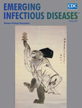
Human minus Three Pieces of Hair [PDF - 216 KB - 2 pages]
| EID | Potter P. Human minus Three Pieces of Hair. Emerg Infect Dis. 2012;18(10):1711-1712. https://doi.org/10.3201/eid1810.ac1810 |
|---|---|
| AMA | Potter P. Human minus Three Pieces of Hair. Emerging Infectious Diseases. 2012;18(10):1711-1712. doi:10.3201/eid1810.ac1810. |
| APA | Potter, P. (2012). Human minus Three Pieces of Hair. Emerging Infectious Diseases, 18(10), 1711-1712. https://doi.org/10.3201/eid1810.ac1810. |


