Volume 18, Number 10—October 2012
Research
Autochthonous and Dormant Cryptococcus gattii Infections in Europe
Cite This Article
Citation for Media
Abstract
Until recently, Cryptococcus gattii infections occurred mainly in tropical and subtropical climate zones. However, during the past decade, C. gattii infections in humans and animals in Europe have increased. To determine whether the infections in Europe were acquired from an autochthonous source or associated with travel, we used multilocus sequence typing to compare 100 isolates from Europe (57 from 40 human patients, 22 from the environment, and 21 from animals) with 191 isolates from around the world. Of the 57 human patient isolates, 47 (83%) were obtained since 1995. Among the 40 patients, 24 (60%) probably acquired the C. gattii infection outside Europe; the remaining 16 (40%) probably acquired the infection within Europe. Human patient isolates from Mediterranean Europe clustered into a distinct genotype with animal and environmental isolates. These results indicate that reactivation of dormant C. gattii infections can occur many years after the infectious agent was acquired elsewhere.
During the past decade, the basidiomycetous yeast Cryptococcus gattii has stepped out of the shadows of its sibling C. neoformans. The latter species mainly infects immunocompromised persons, and C. gattii mainly infects apparently immunocompetent persons. C. neoformans is found globally, and C. gattii has been mostly limited to tropical and subtropical areas in Central Africa, northern Australia, and Central and South America (1). However, this distribution pattern changed after an unprecedented outbreak of C. gattii emerged in the temperate climate of British Columbia, Canada, and expanded to the Pacific Northwest region of Canada and the United States (1,2). Epidemiologic studies have shown that C. gattii occurs in areas other than tropical or subtropical zones, such as in Mediterranean Europe, northern Europe, and western Australia (3–5).
For the purpose of studying the epidemiology of C. gattii, a broad variety of molecular biological techniques have been developed, including PCR fingerprinting, restriction fragment length polymorphism analysis of the PLB1 and URA5 loci, amplified fragment length polymorphism (AFLP) fingerprint analysis, and several multilocus sequence typing (MLST) approaches (6–9). These laboratory investigations have shown that C. gattii can be divided into 5 distinct genotypes: AFLP4/VGI, AFLP5/VGIII, AFLP6/VGII, AFLP7/VGIV, and AFLP10/VGIV (8,9). Serotype B strains occur in genotypes AFLP4/VGI, AFLP6/VGII, and AFLP10/VGIV; serotype C strains are restricted to genotypes AFLP5/VGIII and AFLP7/VGIV (8).
Recently, a consensus MLST scheme was proposed for epidemiologic investigations of C. gattii and C. neoformans, specifically, the nuclear loci CAP59, GPD1, IGS1, LAC1, PLB1, SOD1, and URA5 (9). So far, this consensus MLST scheme has been used to study the population structure of C. neoformans strains from Thailand and C. gattii strains from Australia (3,10).
We investigated the occurrence of C. gattii in Europe, focusing on whether this pathogen is emerging and, if so, how to explain this emergence pattern. Furthermore, we explored whether the infections originated from Europe or were introduced from other continents. To achieve these goals, members of the European Confederation of Medical Mycology were asked to send recently obtained human patient isolates of the species for detailed AFLP and MLST analyses. Thus, the genetic diversity of the yeast was used to trace its geographic origin to identify where the infections were acquired. AFLP genotyping results were combined with published C. gattii MLST results from Byrnes et al. (11) and Fraser et al. (12), which were extended to match the Cryptococcus consensus MLST scheme (9). Our study produced the following 5 conclusions: all hitherto known genotypes of C. gattii are emerging in Europe; genotype AFLP4/VGI isolates predominate; a C. gattii cluster, which is endemic to Mediterranean Europe and genetically distinct from the other populations, exists; several human infections are caused by travel-related acquisition of C. gattii outside Europe; and autochthonous cases occur in Europe.
Strains and Media
We compared a collection of 107 isolates collected from Europe with 194 isolates collected globally (Table 1; Technical Appendix Table 1). All isolates were checked for purity and cultivated on malt extract agar medium (Oxoid, Basingstoke, UK). Cultures were incubated for 2 days at 30°C. A working collection was made by growing C. gattii strains on malt extract agar slants for 2 days at 30°C, after which the strains were stored at 4°C. Strains were stored long term at −80°C by using the Microbank system (Pro-Lab Diagnostics, Richmond Hill, Ontario, Canada).
Amplification and Sequencing of MLST Loci
Genomic DNA extraction, AFLP genotyping, and mating-type determination by amplification of either the STE12a or STE12α locus were performed as described (8). The 7 nuclear consensus MLST loci (CAP59, GPD1, IGS1, LAC1, PLB1, SOD1, and URA5) were amplified by using the preferred primer combinations (9). To compare the current set of C. gattii strains with those from a published C. gattii population biology study, we included the 3 nuclear loci that are not part of the consensus MLST scheme (CAP10, MPD1, and TEF1α) (12). We also included isolates from a global study by Byrnes et al. (11) and Fraser et al. (12) and subjected them to amplification and sequencing of the CAP59, SOD1, and URA5 loci.
Amplifications were conducted in a 25-µL PCR mixture containing 37.5 mmol/L MgCl2 (Bioline, London, UK), 1× PCR buffer (Bioline), 1.9 mmol/L dNTPs (Bioline), 0.5 U Taq DNA polymerase (Bioline), 5 pmol of both primers (Biolegio, Nijmegen, the Netherlands) (Technical Appendix Table 2), and ≈100 ng of genomic DNA. PCRs were conducted with an initial denaturation step at 94°C for 5 min, followed by 35 cycles of denaturation at 94°C for 30s, annealing for 30s (see Technical Appendix Table 2 for optimal annealing temperatures), extension at 72°C for 1 min, followed by 72°C for 5 min and a final dwell at 21°C.
Sequencing reactions were conducted with the BigDye version 3.1 chemistry kit (Applied Biosystems, Foster City, CA, USA) as described (13). For all amplification products except CAP59, the initial amplification primers were used for sequencing reactions. For CAP59, the newly designed forward primer CAP59L-Fwd and the original reverse primer JOHE15438 (12) were used.
Sequence Alignment and Phylogenetic and Recombination Analyses
Consensus sequences were assembled and checked for ambiguities by using SeqMan version 8.0.2 (DNASTAR, Madison, WI, USA). Sequence alignments were generated with MEGA version 5 (14) by using the standard settings and manual correction. The genome sequence databases of reference strains H99 (culture collection no. CBS8710; C. neoformans variety grubii; AFLP1/VNI; Broad Institute [www.broadinstitute.org/annotation/genome/cryptococcus_neoformans/]) and JEC21 (culture collection no. CBS10513; C. neoformans variety neoformans; AFLP2/VNIV; Stanford Genome Technology Center [www-sequence.stanford.edu/group/C.neoformans/]) were used to extract the corresponding sequences for all 10 investigated nuclear loci to serve as an outgroup. The best fitting nucleotide substitution model was determined by using MrModeltest version 2 (15) and was conducted for the complete C. gattii 10-loci MLST, for the accepted consensus MLST scheme (CAP59, GPD1, IGS1, LAC1, PLB1, SOD1, and URA5) (9), and a previously launched C. gattii MLST scheme (CAP10, GPD1, IGS1, LAC1, MPD1, PLB1, and TEF1) (12). As a result, the HKY G+I model (Hasegawa-Kishino-Yano plus gamma distributed with invariant sites) was the best model to use for analyzing the phylogeny of the C. gattii isolates for all 3 datasets. The evolutionary history was inferred by using the maximum-likelihood method in MEGA version 5 (14). A bootstrap consensus tree was inferred from 1,000 replicates to show the relevant lineages obtained in this analysis.
We calculated the haplotype diversity (HR), equal to the Simpson diversity index (D), by using the Microsoft Excel (Microsoft, Redmond, WA, USA) add-in called Haplotype Analysis (16). For this purpose, sequences were collapsed into sequence type numbers (Technical Appendix Table 1).
AFLP Genotypes and Geographic Distribution
The 301 C. gattii isolates collected from Europe and other areas around the world could be divided into the following genotypes: 146 AFLP4/VGI (50.2%; 72 from Europe), 22 AFLP5/VGIII (7.6%; 1 from Europe), 108 AFLP6/VGII (37.1%; 23 from Europe), 13 AFLP7/VGIV (4.5%; 2 from Europe), and 2 AFLP10/VGIV (0.7%; both from Europe). From 10 isolates (7 AFLP8 [5 from Europe] and 3 AFLP9 [2 from Europe]), genotypes represented interspecies C. neoformans × C. gattii hybrids. These 10 isolates were excluded from further analysis because amplified fragments of hybrid isolates will result in mixtures of different alleles. The 57 human patient isolates from Europe were obtained from 40 patients (Technical Appendix Table 1). The genotypic diversity of the remaining 291 C. gattii isolates (100 from Europe, 191 from other areas) with a haploid genotype is shown in the Table.
Phylogeographic Origin
Sequence type diversity was calculated for each of the MLST loci, all 10 loci, and the combined datasets according to Fraser et al. (12) and Meyer et al. (9) (Technical Appendix Table 3). The CAP10 locus showed the lowest overall diversity (nST = 19; DST = 0.765) and the IGS1 locus showed the highest diversity (nST = 52; DST = 0.930). The 10-loci MLST dataset showed the highest diversity (nST = 150; DST = 0.975), followed by the MLST scheme of Meyer et al. (DST = 0.971) (9) and Fraser et al. (DST of 0.959) (12). The latter MLST scheme differentiated more sequence types than the consensus MLST scheme (nST = 136 vs. 127).
Figure
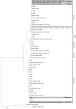
Figure. . . Maximum-likelihood phylogenetic analysis based on 10-loci multilocus sequence type data of Cryptococcus gattii isolates (condensed). Phylogenetic relatedness of 150 STs representing the 291 C. gattii isolates, calculated by using...
Maximum-likelihood analysis of the MLST data showed that the C. gattii isolates clustered in 5 monophyletic clusters, which were highly supported and agreed with the AFLP genotypes (Figure, Technical Appendix Figure). When each genotypic cluster was separately analyzed, support values of the branches were low, <75 (Technical Appendix Figure). For genotype AFLP4/VGI isolates, bootstrap support values for nearly all branches were low; for the second largest group formed by AFLP6/VGII isolates, branches were better supported.
Phylogenetic analysis demonstrated that several human patient genotype AFLP4/VGI isolates from Europe had an autochthonous origin because they formed a separate cluster with environmental and animal isolates from the same area. A set of isolates from human patients on the Iberian Peninsula (CCA232, CCA242L, CCA242T, CCA311, CCA312, CL6148) were found to be genetically indistinguishable from isolates obtained from animals or the environment in the same area (indicated in Technical Appendix Figure as the European Mediterranean cluster). The human patient isolates from Europe with genotype AFLP4/VGI, which were probably acquired within Europe because patients did not have histories of travel outside Europe, are IP2005/215 and IP2006/958 (France), RKI85/888 (Germany), 5UM and 75UM (Italy), RKI97/482 (Portugal), and CBS2502 (the Netherlands). Some of these human patient isolates were closely related (e.g., IP2005/215 and RKI97/482) or even genetically indistinguishable (e.g., CBS2502 and RKI85/888).
A large proportion of the human patient and environmental isolates of C. gattii AFLP4/VGI from Italy and Spain formed a novel autochthonous Mediterranean MLST cluster that was genetically homogeneous, irrespective of their origin or mating type (Figure, Technical Appendix Figure). C. gattii AFLP4/VGI was involved with numerous small outbreaks among goats in Spain, and the isolates were genetically indistinguishable from recently obtained human patient, animal, and environmental isolates from different provinces in Spain as well as from AFLP4/VGI mating-type a isolates from Italy (Technical Appendix Figure).
Several C. gattii genotype AFLP6/VGII infections were found to have originated in Europe (e.g., the IP1998/1037–1 and −2, IP2003/125, and CCA242 isolates), and all fell within a cluster that could not be linked to an environmental source. The same holds true for the human patient isolates from Greece (AV54S, –W, and IUM01–4731), which came from the same patient who had no history of travel outside Greece.
In the phylogenetic analysis, isolates from human patients in Europe were also observed next to those originating from other geographic areas. The most striking example was a set of 4 human patient isolates from citizens of Denmark, the Netherlands, Germany, and Switzerland, in whom cryptococcosis developed after they had visited Vancouver Island, British Columbia, Canada. The isolates obtained from these tourists (CBS10485, RKI06/496, RKI01/774, and CBS10866, respectively) had MLST profiles identical to that of C. gattii AFLP6A/VGIIa, the genotype that caused the Vancouver Island outbreak (Figure, Technical Appendix Figure) (17–19). The outbreak-related sets of AFLP6A/VGIIa and AFLP6C/VGIIc isolates from the Vancouver Island and Pacific Northwest outbreaks, respectively, are within outbreak-specific clusters (Figure, Technical Appendix Figure). A similar finding was observed for a set of 5 human patient C. gattii AFLP6B/VGIIb isolates (IP1996/1120–1 and −2, IP1999/901–1 and −2, and IP2000/87) in France, which had been obtained from patients who had emigrated from Africa to France and which were genetically indistinguishable from an isolate (IP2001/935–1) from a resident of Senegal (Technical Appendix Figure). This set of 5 isolates from France and 1 from Senegal were closely related to isolates from the Vancouver Island and Pacific Northwest outbreaks; however, it seems unlikely that the patient from Senegal had traveled to these outbreak areas, and none of the patients in France reported having traveled to Canada or the United States.
A set of C. gattii AFLP4/VGI mating-type α isolates from Portugal (IP1997/18) and Belgium (IHEM19725B and –S) was found to be indistinguishable from human patient isolates from the Democratic Republic of the Congo and Rwanda (B3939 and CBS6289, which were both mating-type a, and IHEM10602S, IHEM10769S, and IHEM10769W, which were mating-type α) (Figure, Technical Appendix Figure). Both phenotypically different isolates of IHEM19725 were obtained from an HIV-infected patient from Rwanda who had emigrated to Belgium. The CBS1622 isolate from Europe, obtained from a patient with the oldest documented case of a C. gattii infection (20), was found to be genetically indistinguishable from a set of isolates from North America.
The single C. gattii AFLP5/VGIII isolate from a 24-year-old immunocompetent patient from Germany (isolate RKI97/310) (21) clustered with human patient and environmental isolates from Mexico (Technical Appendix Figure). The 2 C. gattii AFLP7/VGIV isolates CBS7952D and CBS7952S, obtained from an HIV-infected patient in Sweden, were genetically indistinguishable from each other and had a unique MLST profile that clustered with human patient isolates from Africa. According to the patient’s history, years before the onset of cryptococcal infection, she had emigrated from Zambia to Sweden (Technical Appendix Figure). Another example of a reactivated dormant C. gattii infection is that of the human patient isolate from Italy, IUM92–6682 (AFLP4/VGI), obtained from an immunocompetent immigrant from Brazil, which had an identical MLST profile to 6 human patient mating-type α isolates (IHEM14934, IHEM14956, IHEM14965, IHEM14968, IHEM14976, and IHEM14984) from Brazil (Figure, Technical Appendix Figure).
Infection was acquired outside Europe for 24 of these patients (31 isolates) and within Europe for 16 patients (26 isolates) (Table, Technical Appendix Figure). Among these 57 human patient isolates, most (47 [82.5%]) were obtained since 1995, the remaining 10 (17.5%) were isolated during 1895–1994. One of these isolates originated from 1985, another 2 isolates (CBS1622 and CBS2502), from 1895 and 1957, were retrospectively found to represent C. gattii isolates from Europe (20,22).
In Europe, C. gattii has been reported as a rare cause of apparently autochthonous cryptococcal infections (1). The earliest documented case that turned out to be caused by C. gattii in Europe was made by Curtis in 1896 (20). Until the 1980s, cryptococcosis was rarely observed in Europe and C. gattii infections were especially rare; only 2 cases have retrospectively been found (20,22). In the 1980s, infections were reported for 2 immunocompetent citizens of Germany, who had never traveled abroad (23,24). Subsequent case reports described C. gattii infections that were imported or acquired in Europe (25,26). Since 1995, the number of reports of C. gattii infections increased and describe C. gattii infections in humans and animals from Greece (27), Italy (28,29), and Spain (5,30–33). In the current study, 8 (20%) cases were observed until 1995, and 32 (80%) cases in humans have been observed since 1995. We excluded cases for which an isolate was not available for confirmation. This exclusion is especially relevant in that we re-identified a reported C. gattii infection as actually being caused by C. neoformans (34). These data suggest that during the past 2 decades, C. gattii has been emerging in Europe.
In the current study, 16 (40%) of the human patient isolates from Europe were found to have an autochthonous origin in Europe that either could be linked to environmental isolates from the Mediterranean area or that came from patients who had never traveled outside their resident country. A total of 24 (60%) isolates could be linked to C. gattii–endemic regions in Brazil, the United States, Africa, and the Vancouver Island outbreak region. These observations demonstrate that C. gattii infections can be imported subclinically and can cause infections after being dormant for many years.
Nearly all isolates from Mediterranean Europe belonged to genotype AFLP4/VGI, and MLST analysis showed that these isolates form a separate cluster within this genotype (Figure, Technical Appendix Figure) (5). C. gattii genotype AFLP4/VGI has also recently been reported from the environment in the Netherlands, but these isolates were not similar to any of the AFLP4/VGI isolates from Mediterranean Europe or to isolate CBS2502, which was isolated in 1957 from a pregnant citizen of the Netherlands, who had never traveled abroad but who died of cryptococcosis (4). Isolate CBS2502 is genotypically identical to isolate RKI85/888, which was isolated from a previously healthy citizen of Germany, who also had never traveled outside Germany. These 2 cases strongly suggest that different genotypes of C. gattii AFLP4/VGI occur in the environment of northwestern Europe because isolates from patients were genetically different from the recently reported isolates from the environment of the Netherlands (this study; 4).
In conclusion, C. gattii is emerging in Europe, and the isolates from Europe can be divided into 5 genotypic clusters. Most C. gattii infections in Europe are probably autochthonous, and several infections are proven to have been acquired outside the European continent, e.g., during visits to C. gattii–endemic regions, such as the Vancouver Island outbreak area or before migration to Europe from C. gattii–endemic regions in Africa and South America. Reactivation of dormant C. gattii infections and of infections acquired outside Europe after immune suppression occurs more often than previously assumed. This finding might suggest that C. gattii infections caused by certain genotypes are associated with altered immune status of the human host. Thus, C. gattii is probably a more opportunistic pathogen, as has recently been hypothesized, than a strictly primary pathogen (18,35). C. gattii isolates with genotypes AFLP5/VGIII and AFLP7/VGIV were rarely found in Europe and were all acquired outside the European continent. However, these genotypes have frequently been isolated from HIV-infected persons and other immunocompromised patients in Africa and the American continents (1,7,36). A connection seems to exist between these C. gattii genotypes and the host’s immune status. Further epidemiologic and immunologic research is needed to unravel this apparent correlation.
Dr Hagen is a medical molecular microbiologist trainee at the Canisius-Wilhelmina Hospital in Nijmegen, the Netherlands. His research interests include medical mycology, especially the development of novel molecular approaches to understand the epidemiology of fungal pathogens.
Acknowledgments
We thank Karen Bartlett, Alf Botha, Eddy Byrnes III, Manuel Cuenca-Estrella, Françoise Dromer, Martine Gari-Toussaint, Jesús Guinea, Joseph Heitman, Connie Jarstrand, Margareth Ip, Greetje Kampinga, Maarten Scholing, and Marianna Viviani for providing Cryptococcus spp. isolates.
This work was conducted at the CBS-KNAW Fungal Biodiversity Centre in Utrecht, the Netherlands. The data are available online (www.cbs.knaw.nl/publications/online.aspx). Research performed by F.H. was financially supported by the Odo van Vloten Foundation, the Netherlands.
References
- Springer DJ, Chaturvedi V. Projecting global occurrence of Cryptococcus gattii. Emerg Infect Dis. 2010;16:14–20. DOIPubMedGoogle Scholar
- Kidd SE, Hagen F, Tscharke RL, Huynh M, Bartlett KH, Fyfe M, A rare genotype of Cryptococcus gattii caused the cryptococcosis outbreak on Vancouver Island (British Columbia, Canada). Proc Natl Acad Sci U S A. 2004;101:17258–63. DOIPubMedGoogle Scholar
- Carriconde F, Gilgado F, Arthur I, Ellis D, Malik R, van de Wiele N, Clonality and α-a recombination in the Australian Cryptococcus gattii VGII population—an emerging outbreak in Australia. PLoS One. 2011;6:e16936. DOIPubMedGoogle Scholar
- Chowdhary A, Randhawa HS, Boekhout T, Hagen F, Klaassen CHW, Meis JF. Temperate climate niche for Cryptococcus gattii in northern Europe. Emerg Infect Dis. 2012;18:172–4. DOIPubMedGoogle Scholar
- Colom MF, Hagen F, Gonzalez A, Mellado A, Morera N, Linares C, Ceratonia siliqua (carob) trees as natural habitat and source of infection by Cryptococcus gattii in the Mediterranean environment. Med Mycol. 2012;50:67–73.PubMedGoogle Scholar
- Boekhout T, Theelen B, Diaz M, Fell JW, Hop WC, Abeln EC, Hybrid genotypes in the pathogenic yeast Cryptococcus neoformans. Microbiology. 2001;147:891–907.PubMedGoogle Scholar
- Bovers M, Hagen F, Boekhout T. Diversity of the Cryptococcus neoformans–Cryptococcus gattii species complex. Rev Iberoam Micol. 2008;25:S4–12. DOIPubMedGoogle Scholar
- Hagen F, Illnait-Zaragozi MT, Bartlett KH, Swinne D, Geertsen E, Klaassen CH, In vitro antifungal susceptibilities and amplified fragment length polymorphism genotyping of a worldwide collection of 350 clinical, veterinary, and environmental Cryptococcus gattii isolates. Antimicrob Agents Chemother. 2010;54:5139–45. DOIPubMedGoogle Scholar
- Meyer W, Aanensen DM, Boekhout T, Cogliati M, Diaz MR, Esposto MC, Consensus multi-locus sequence typing scheme for Cryptococcus neoformans and Cryptococcus gattii. Med Mycol. 2009;47:561–70. DOIPubMedGoogle Scholar
- Simwami SP, Khayhan K, Henk DA, Aanensen DM, Boekhout T, Hagen F, Low diversity Cryptococcus neoformans variety grubii multilocus sequence types from Thailand are consistent with an ancestral African origin. PLoS Pathog. 2011;7:e1001343. DOIPubMedGoogle Scholar
- Byrnes EJ III, Li W, Lewit Y, Ma H, Voelz K, Ren P, Emergence and pathogenicity of highly virulent Cryptococcus gattii genotypes in the northwest United States. PLoS Pathog. 2010;6:e1000850. DOIPubMedGoogle Scholar
- Fraser JA, Giles SS, Wenink EC, Geunes-Boyer SG, Wright JR, Diezmann S, Same-sex mating and the origin of the Vancouver Island Cryptococcus gattii outbreak. Nature. 2005;437:1360–4. DOIPubMedGoogle Scholar
- Bovers M, Hagen F, Kuramae EE, Boekhout T. Six monophyletic lineages identified within Cryptococcus neoformans and Cryptococcus gattii by multi-locus sequence typing. Fungal Genet Biol. 2008;45:400–21. DOIPubMedGoogle Scholar
- Tamura K, Peterson D, Peterson N, Stecher G, Nei M, Kumar S. MEGA5: Molecular Evolutionary Genetics Analysis using maximum likelihood, evolutionary distance, and maximum parsimony methods. Mol Biol Evol. 2011;28:2731–9. DOIPubMedGoogle Scholar
- Nylander JAA. MrModeltest v.2. Uppsala: Evolutionary Biology Centre, Uppsala University: 2004 [cited 2012 Aug 9]. http://www.abc.se/~nylander/
- Eliades N-G, Eliades DG. Haplotype Analysis: software for analysis of haplotype data. Forest Goettingen (Germany): Genetics and Forest Tree Breeding, Georg-August University Goettingen,. 2009 [cited 2012 Aug 9]. http://www.uni-goettingen.de/en/134935.html
- Georgi A, Schneemann M, Tintelnot K, Calligaris-Maibach RC, Meyer S, Weber R, Cryptococcus gattii meningoencephalitis in an immunocompetent person 13 months after exposure. Infection. 2009;37:370–3. DOIPubMedGoogle Scholar
- Hagen F, van Assen S, Luijckx GJ, Boekhout T, Kampinga GA. Activated dormant Cryptococcus gattii infection in a Dutch tourist who visited Vancouver Island (Canada): a molecular epidemiological approach. Med Mycol. 2010;48:528–31. DOIPubMedGoogle Scholar
- Lindberg J, Hagen F, Laursen A, Stenderup J, Boekhout T. Cryptococcus gattii risk for tourists visiting Vancouver Island, Canada. Emerg Infect Dis. 2007;13:178–9. DOIPubMedGoogle Scholar
- Curtis F. Contribution a l’étude de la saccharomycose humaine. Ann Inst Pasteur (Paris). 1896;10:449–68.
- Grosse P, Tintelnot K, Söllner O, Schmitz B. Encephalomyelitis due to Cryptococcus neoformans var. gattii presenting as spinal tumour: case report and review of the literature. J Neurol Neurosurg Psychiatry. 2001;70:113–6. DOIPubMedGoogle Scholar
- Janssens J, Beetstra A. Torulosis in a pregnant female, with localization in the lungs [in Dutch]. Ned Tijdschr Geneeskd. 1957;101:824–6.PubMedGoogle Scholar
- Kohl KH, Hof H, Schrettenbrunner A, Seeliger HP, Kwon-Chung KJ. Cryptococcus neoformans var. gattii in Europe. Lancet. 1985;325:1515. DOIPubMedGoogle Scholar
- Schaberg T, Mai J, Thalmann U, Seibold M, Staib F. Cryptococcoma of the lung—a contribution to diagnosis and therapy [in German]. Internist (Berl). 1988;29:510–5.PubMedGoogle Scholar
- Dromer F, Ronin O, Dupont B. Isolation of Cryptococcus neoformans var. gattii from an Asian patient in France: evidence for dormant infection in healthy subjects. J Med Vet Mycol. 1992;30:395–7. DOIPubMedGoogle Scholar
- Dromer F, Mathoulin S, Dupont B, Laporte A. Epidemiology of cryptococcosis in France: a 9-year survey (1985–1993). French Cryptococcosis Study Group. Clin Infect Dis. 1996;23:82–90. DOIPubMedGoogle Scholar
- Velegraki A, Kiosses VG, Pitsouni H, Toukas D, Daniilidis VD, Legakis NJ. First report of Cryptococcus neoformans var. gattii serotype B from Greece. Med Mycol. 2001;39:419–22.PubMedGoogle Scholar
- Iatta R, Hagen F, Fico C, Lopatriello N, Boekhout T, Montagna MT. Cryptococcus gattii infection in an immunocompetent patient from southern Italy. Mycopathologia. 2012 ;174:87–92. DOIPubMedGoogle Scholar
- Montagna MT, Viviani MA, Pulito A, Aralla C, Tortorano AM, Fiore L, Cryptococcus neoformans var. gattii in Italy. Note II. Environmental investigation related to an autochthonous clinical case in Apulia. J Mycol Med. 1997;7:93–6.
- Baró T, Torres-Rodríguez JM, De Mendoza MH, Morera Y, Alía C. First identification of autochthonous Cryptococcus neoformans var. gattii isloated from goats with predominantly severe pulmonary disease in Spain. J Clin Microbiol. 1998;36:458–61.PubMedGoogle Scholar
- Colom MF, Frasés S, Ferrer C, Jover A, Andreu M, Reus S, First case of human cryptococcosis due to Cryptococcus neoformans var. gattii in Spain. J Clin Microbiol. 2005;43:3548–50. DOIPubMedGoogle Scholar
- Guinea J, Hagen F, Peláez T, Boekhout T, Tahoune H, Torres-Narbona M, Antifungal susceptibility, serotyping, and genotyping of clinical Cryptococcus neoformans isolates collected during 18 years in a single institution in Madrid, Spain. Med Mycol. 2010;48:942–8. DOIPubMedGoogle Scholar
- Solla I, Morano LE, Vasallo F, Cuenca-Estrella M. Cryptococcus gattii meningitis: observation in a Spanish patient [in Spanish]. Enferm Infecc Microbiol Clin. 2008;26:395–6. DOIPubMedGoogle Scholar
- Bodasing N, Seaton RA, Shankland GS, Kennedy D. Cryptococcus neoformans var. gattii meningitis in an HIV-positive patient: first observation in the United Kingdom. J Infect. 2004;49:253–5. DOIPubMedGoogle Scholar
- MacDougall L, Fyfe M, Romney M, Starr M, Galanis E. Risk factors for Cryptococcus gattii infection, British Columbia, Canada. Emerg Infect Dis. 2011;17:193–9. DOIPubMedGoogle Scholar
- Litvintseva AP, Thakur R, Reller LB, Mitchell TG. Prevalence of clinical isolates of Cryptococcus gattii serotype C among patients with AIDS in sub-Saharan Africa. J Infect Dis. 2005;192:888–92. DOIPubMedGoogle Scholar
Figure
Table
Cite This ArticleTable of Contents – Volume 18, Number 10—October 2012
| EID Search Options |
|---|
|
|
|
|
|
|
Please use the form below to submit correspondence to the authors or contact them at the following address:
Ferry Hagen, Canisius-Wilhelmina Hospital, Department of Medical Microbiology and Infectious Diseases, Weg door Jonkerbos 100, NL-6532 SZ Nijmegen, the Netherlands
Top