Volume 18, Number 2—February 2012
Dispatch
Shuni Virus as Cause of Neurologic Disease in Horses
Charmaine van Eeden, June H. Williams, Truuske G.H. Gerdes, Erna van Wilpe, Adrianne Viljoen, Robert Swanepoel, and Marietjie Venter
Figure 1
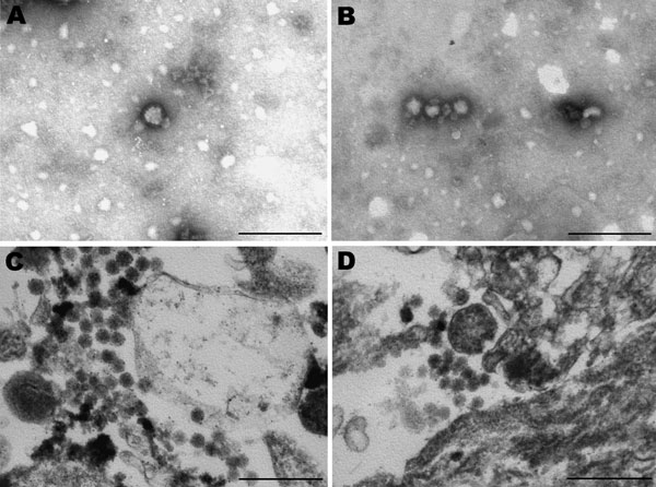
Figure 1. Electron micrographs of Vero cells infected with virus from horse SAE 18/09. A, B) Negative stain showing fringed particles (bunyavirus size) with bleb formation. C, D) Resin section showing spherical and pleomorphic bunyavirus particles in the range of 80–100 nm. Scale bars = 250 nm.
Page created: January 19, 2012
Page updated: January 19, 2012
Page reviewed: January 19, 2012
The conclusions, findings, and opinions expressed by authors contributing to this journal do not necessarily reflect the official position of the U.S. Department of Health and Human Services, the Public Health Service, the Centers for Disease Control and Prevention, or the authors' affiliated institutions. Use of trade names is for identification only and does not imply endorsement by any of the groups named above.