Volume 18, Number 5—May 2012
CME ACTIVITY - Research
Risk Factors for Intestinal Invasive Amebiasis in Japan, 2003–2009
Naoyoshi Nagata , Takuro Shimbo, Junichi Akiyama, Ryo Nakashima, So Nishimura, Tomoyuki Yada, Koji Watanabe, Shinichi Oka, and Naomi Uemura
Figure 1
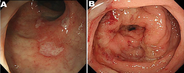
Figure 1. . . Endoscopic features of amebic colitis, Japan, 2003–2009. A) Colonoscopy showing ulcers in the rectum. B) Colonoscopy showing multiple erosions with exudates surrounded by edematous mucosa in the sigmoid colon.
Page created: April 16, 2012
Page updated: April 16, 2012
Page reviewed: April 16, 2012
The conclusions, findings, and opinions expressed by authors contributing to this journal do not necessarily reflect the official position of the U.S. Department of Health and Human Services, the Public Health Service, the Centers for Disease Control and Prevention, or the authors' affiliated institutions. Use of trade names is for identification only and does not imply endorsement by any of the groups named above.