Skip directly to site content Skip directly to page options Skip directly to A-Z link Skip directly to A-Z link Skip directly to A-Z link
Volume 19, Number 2—February 2013
Letter

Delayed Diagnosis of Dirofilariasis and Complex Ocular Surgery, Russia

Boris Ilyasov, Vladimir Kartashev, Nikolay Bastrikov, Rodrigo Morchón, Javier González-Miguel, and Fernando SimónComments to Author 
Author affiliations: Author affiliations: Rostov Oblast Diagnostic Centre, Rostov-na-Donu, Russia (B. Ilyasov); Rostov State Medical University, Rostov-na-Donu (V. Kartashev); Hospital of the North Caucasus Branch of the Russian Railway, Rostov-na-Donu (N. Bastrikov); University of Salamanca, Salamanca, Spain (R. Morchón, J. González-Miguel, F. Simón)

Main Article

Figure

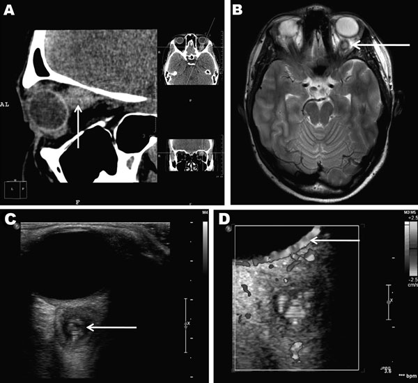
Retroocular nodule of a Dirofilaria repens worm detected in a 20-year-old woman, Rostov-na-Donu, Russia. The cyst (arrows) is shown by computed tomography scan (A) and magnetic resonance imaging (B). Ultrasonography images (C) show a worm-like structure inside the cyst (arrow), and color Doppler imaging (D) shows marginal vascularization of the lesion).

Figure. . Retroocular nodule of a Dirofilaria repens worm detected in a 20-year-old woman, Rostov-na-Donu, Russia. The cyst (arrows) is shown by computed tomography scan (A) and magnetic resonance imaging (B). Ultrasonography image (C) shows a worm-like structure inside the cyst (arrow), and color Doppler imaging (D) shows marginal vascularization of the lesion).

Main Article

Page created: January 23, 2013
Page updated: January 23, 2013
Page reviewed: January 23, 2013
The conclusions, findings, and opinions expressed by authors contributing to this journal do not necessarily reflect the official position of the U.S. Department of Health and Human Services, the Public Health Service, the Centers for Disease Control and Prevention, or the authors' affiliated institutions. Use of trade names is for identification only and does not imply endorsement by any of the groups named above.
file_external