Volume 19, Number 3—March 2013
Letter
Armillifer armillatus Pentastomiasis in African Immigrant, Germany
Dennis Tappe , Alexandra Haeupler, Hansjörg Schäfer, Paul Racz, Jakob P. Cramer, and Sven Poppert
Figure
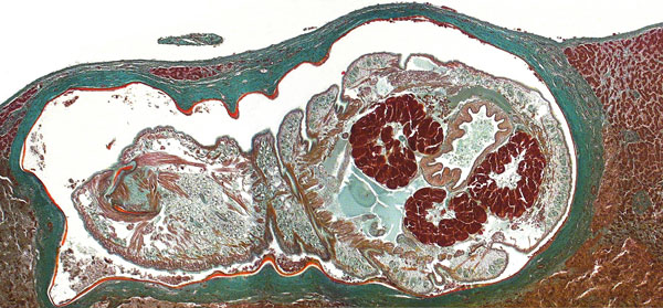
Figure. . . Oblique cross-section of liver of a patient (immigrant) from Togo, showing a well-preserved Armillifer armillatus nymph in a subcapsular location. The annulated parasite is encapsulated by its shed cuticle (exuvia) and dense fibrosis. Consistent with the viable type of a pentastomid lesion (3), no inflammatory infiltrate is visible. This image also shows internal structures of the pentastome, such as prominent bunches of acidophilic glands surrounding the intestine (Masson’s trichrome stain, original magnification ×10).
References
- Latif B, Omar E, Heo CC, Othman N, Tappe D. Human pentastomiasis caused by Armillifer moniliformis in Malaysian Borneo. Am J Trop Med Hyg. 2011;85:878–81. DOIPubMedGoogle Scholar
- Tappe D, Meyer M, Oesterlein A, Jaye A, Frosch M, Schoen C, Transmission of Armillifer armillatus ova at snake farm, The Gambia, West Africa. Emerg Infect Dis. 2011;17:251–4. DOIPubMedGoogle Scholar
- Tappe D, Büttner DW. Diagnosis of human visceral pentastomiasis. PLoS Negl Trop Dis. 2009;3:e320. DOIPubMedGoogle Scholar
- Lavarde V, Fornes P. Lethal infection due to Armillifer armillatus (Porocephalida): a snake-related parasitic disease. Clin Infect Dis. 1999;29:1346–7. DOIPubMedGoogle Scholar
- Guardia SN, Sepp H, Scholten T, Morava-Protzner I. Pentastomiasis in Canada. Arch Pathol Lab Med. 1991;115:515–7.PubMedGoogle Scholar
- Cagnard V, Nicolas-Randegger J, Dago Akribi A, Rain B, Nozais JP, Essoh Nomel P, Generalized and lethal pentastomiasis due to Armillifer grandis (Hett, 1915) [in French]. Bull Soc Pathol Exot. 1979;72:345–52.PubMedGoogle Scholar
- Brookins MD, Wellehan JF, Roberts JF, Allison K, Curran SS, Childress AL, Massive visceral pentastomiasis caused by Porocephalus crotali in a dog. Vet Pathol. 2009;46:460–3. DOIPubMedGoogle Scholar
- Prathap K, Lau KS, Bolton JM. Pentastomiasis: a common finding at autopsy among Malaysian aborigines. Am J Trop Med Hyg. 1969;18:20–7 .PubMedGoogle Scholar
- Smith JA, Oladiran B, Lagundoye SB. Pentastomiasis and malignancy. Ann Trop Med Parasitol. 1975;69:503–12.
- Tiendrebeogo H, Levy D, Schmidt D. Human pentastomiasis in Abidjan. A report on 29 cases [in French]. Rev Fr Mal Respir. 1982;10:351–8 .PubMedGoogle Scholar
Page created: January 31, 2013
Page updated: January 31, 2013
Page reviewed: January 31, 2013
The conclusions, findings, and opinions expressed by authors contributing to this journal do not necessarily reflect the official position of the U.S. Department of Health and Human Services, the Public Health Service, the Centers for Disease Control and Prevention, or the authors' affiliated institutions. Use of trade names is for identification only and does not imply endorsement by any of the groups named above.