Novel Mycobacterium tuberculosis Complex Isolate from a Wild Chimpanzee
Mireia Coscolla, Astrid Lewin, Sonja Metzger, Kerstin Maetz-Rennsing, Sébastien Calvignac-Spencer, Andreas Nitsche, Pjotr Wojtek Dabrowski, Aleksandar Radonic, Stefan Niemann, Julian Parkhill, Emmanuel Couacy-Hymann, Julia Feldman, Iñaki Comas, Christophe Boesch, Sebastien Gagneux
1, and Fabian H. Leendertz
1
Author affiliations: Swiss Tropical and Public Health Institute, Basel, Switzerland (M. Coscolla, J. Feldman, S. Gagneux); University of Basel, Basel (M. Coscolla, J. Feldman, S. Gagneux); Centro Superior de Investigación en Salud Pública, Valencia, Spain (M. Coscolla, I. Comas); Robert Koch-Institut, Berlin, Germany (A. Lewin, S. Metzger, S. Calvignac-Spencer, A. Nitsche, P. Wojtek Dabrowski, A. Radonic, F.H. Leendertz); Max-Planck-Institute for Evolutionary Anthropology, Leipzig, Germany (S. Metzger, C. Boesch); German Primate Center, Goettingen, Germany (K. Maetz-Rennsing); Research Centre Borstel, Borstel, Germany (S. Niemann); Wellcome Trust Sanger Institute, Cambridge, UK (J. Parkhill); Laboratoire Nationale de la Pathologie Animale, Bingerville, Côte d’Ivoire (E. Couacy-Hymann); CIBER in Epidemiology and Public Health, Barcelona, Spain (I. Comas)
Main Article
Figure 2
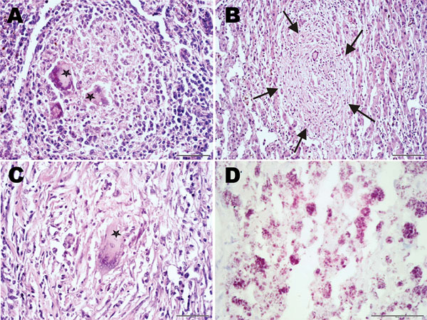
Figure 2. . Histopathologic examination of tissue samples from adult female chimpanzee that was found dead in Taï National Park, Côte d’Ivoire, on August 5, 2009. A) Hematoxylin and eosin (H&E) stain of the spleen shows focal granulomatous inflammation with central accumulation of multinucleated Langhans giant cells (stars). B, C) H&E stain of the liver shows focal granulomatous inflammation within liver parenchyma (B, arrows) and large granulomatous alteration demarcated by fibrous connective tissue infiltrated by Langhans giant cells (C). D) Ziehl-Neelsen stain of the liver shows aggregates of acid-fast bacilli within a large granuloma. Results were consistent with Mycobacterium tuberculosis complex infection.
Main Article
Page created: May 20, 2013
Page updated: May 20, 2013
Page reviewed: May 20, 2013
The conclusions, findings, and opinions expressed by authors contributing to this journal do not necessarily reflect the official position of the U.S. Department of Health and Human Services, the Public Health Service, the Centers for Disease Control and Prevention, or the authors' affiliated institutions. Use of trade names is for identification only and does not imply endorsement by any of the groups named above.