Volume 19, Number 6—June 2013
Synopsis
Cell Culture and Electron Microscopy for Identifying Viruses in Diseases of Unknown Cause
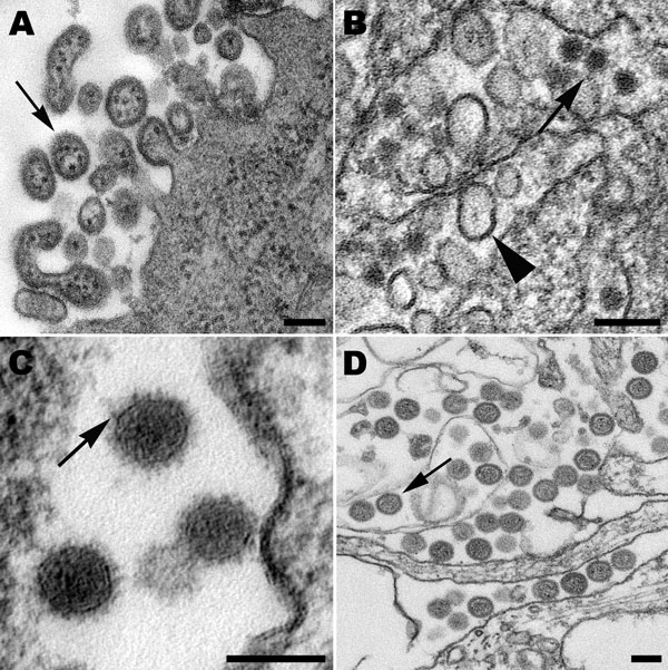
Figure 2

Figure 2. . . A) Extracellular lymphocytic choriomeningitis virus particles (arrow) containing cellular ribosomes. B) West Nile virus particles (arrow) in the lumen of the rough endoplasmic reticulum of an infected cell. Also in the cisternae are smooth membrane vesicles (arrowhead). C) High magnification of Cache Valley virus particles within a Golgi vesicle, showing small surface projections (arrow). D) Extracellular, spherical Homeland virus particles (arrow) with a slightly granular core. Scale bars = 100 nm.
Page created: May 20, 2013
Page updated: May 20, 2013
Page reviewed: May 20, 2013
The conclusions, findings, and opinions expressed by authors contributing to this journal do not necessarily reflect the official position of the U.S. Department of Health and Human Services, the Public Health Service, the Centers for Disease Control and Prevention, or the authors' affiliated institutions. Use of trade names is for identification only and does not imply endorsement by any of the groups named above.