Volume 2, Number 1—January 1996
Synopsis
Emergence of the Ehrlichioses as Human Health Problems
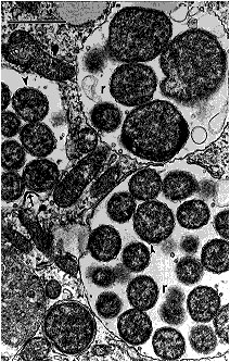
Figure 3

Figure 3. Human monocytic ehrlichiae (Ehrlichia chaffeensis, Sapulpa strain) in host-cell membrane-limited parasitophorous vacuoles (morulae) of a DH82 cell (canine macrophage cell line). Ehrlichial reticulate cells (r) are limited by two membranes. The outer one -- the cell wall membrane -- is usually wavy (arrowheads). One Ehrlichia is dividing by binary fussion (arrow). Bar = 1 µm; magnification, x 18,000. (Courtesy of Vsevolod Popov, University of Texas Medical Branch at Galveston.)
Page created: December 20, 2010
Page updated: December 20, 2010
Page reviewed: December 20, 2010
The conclusions, findings, and opinions expressed by authors contributing to this journal do not necessarily reflect the official position of the U.S. Department of Health and Human Services, the Public Health Service, the Centers for Disease Control and Prevention, or the authors' affiliated institutions. Use of trade names is for identification only and does not imply endorsement by any of the groups named above.