Volume 20, Number 2—February 2014
Dispatch
Lethal Factor and Anti-Protective Antigen IgG Levels Associated with Inhalation Anthrax, Minnesota, USA
Mark D. Sprenkle , Jayne Griffith, William Marinelli, Anne E. Boyer, Conrad P. Quinn, Nicki T. Pesik, Alex Hoffmaster, Joseph Keenan, Billie A. Juni, and David D. Blaney
Figure 1
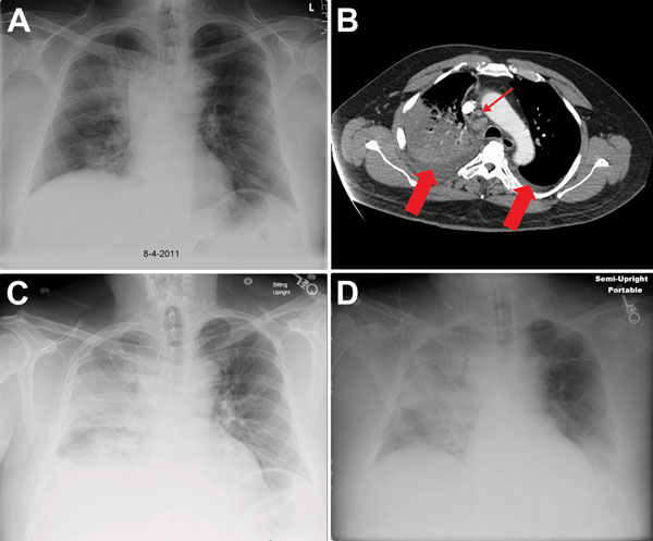
Figure 1. . Chest x-ray and computed tomographic scan images for a patient with inhalation anthrax, Minnesota, USA. A) On hospital day 1, the x-ray image revealed a right upper lobe infiltrate and widening of the mediastinum. B) On hospital day 2, computed tomographic scan of the chest with intravenous contrast showed dense consolidation of the right upper lobe, mediastinal adenopathy (small arrow), and bilateral pleural effusions (large arrows). C) By hospital day 4, progressive infiltrates in the right lung were present. D) By day 6, an increasing left pleural effusion was evident.
Page created: January 17, 2014
Page updated: January 17, 2014
Page reviewed: January 17, 2014
The conclusions, findings, and opinions expressed by authors contributing to this journal do not necessarily reflect the official position of the U.S. Department of Health and Human Services, the Public Health Service, the Centers for Disease Control and Prevention, or the authors' affiliated institutions. Use of trade names is for identification only and does not imply endorsement by any of the groups named above.