Volume 20, Number 2—February 2014
Letter
Laboratory-acquired Buffalopox Virus Infection, India
Figure

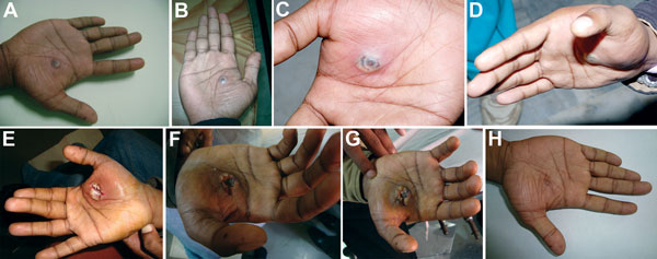
Figure. . Progression of lesion on palm of researcher caused by buffalopox virus infection, India. A) Small vesicle on postinjury day 5. B) Pustule with a central area of necrosis on postinjury day 7. C) Pustule with edema on postinjury day 9. D) Increase in edema on postinjury day 10 (cyanosis is not apparent). E) Lesion after surgical excision on postinjury day 11. F) Lesion on postinjury day 19 (healing was erratic). G) Blackening and thickening around surgical site on postinjury day 30, which then extended to a wider circumference. H) Healed lesion with a black eschar on postinjury day 85.
1These authors contributed equally to this article.
Page created: January 17, 2014
Page updated: January 17, 2014
Page reviewed: January 17, 2014
The conclusions, findings, and opinions expressed by authors contributing to this journal do not necessarily reflect the official position of the U.S. Department of Health and Human Services, the Public Health Service, the Centers for Disease Control and Prevention, or the authors' affiliated institutions. Use of trade names is for identification only and does not imply endorsement by any of the groups named above.