Volume 20, Number 2—February 2014
Dispatch
Replicative Capacity of MERS Coronavirus in Livestock Cell Lines
Figure 2
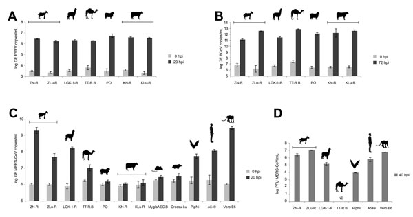
Figure 2. . Analysis of the replication of Rift Valley fever virus (RVFV) clone 13 (A), bovine coronavirus (BCoV) (B), and Middle East respiratory syndrome coronavirus (MERS-CoV) (C) and of the production of infectious MERS-CoV particles (D) in cell lines derived from livestock and peridomestic small mammals on the Arabian Peninsula. Cell lines of human, bat, and primate origin were used as controls. Replication levels for each virus used are given as log of the genome equivalents (GEs) (A–C) or as plaque-forming units (PFUs). Vertical bars indicate ranges; horizontal bars indicate cell line origins. Using panel C as a reference, symbols represent (left to right) goat, alpaca, Arabian camel, sheep, cattle, bank vole, shrew, bat, human, and African green monkey. ND, not detected; hpi, hours postincubation.