Volume 20, Number 4—April 2014
Dispatch
Cetacean Morbillivirus in Coastal Indo-Pacific Bottlenose Dolphins, Western Australia
Figure 1
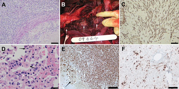
Figure 1. . . . Images of tissue samples from 2 stranded coastal Indo-Pacific bottlenose dolphins (Tursiops aduncus) from Western Australia, Australia. A) Brain of dolphin 2 showing cerebral hemisphere with focally extensive suppurative and necrotizing encephalitis surrounding an arteriole. There are intramural and perivascular septate branching hyphae. Hematoxylin and eosin stain. Scale bar = 50 μm. B) Lung of dolphin 3 showing a transected lobar surface exhibiting multifocal pyogranulomas (white arrows). C) Lung of dolphin 3 showing bronchointerstitial pneumonia with branching septate hyphae within a bronchiolar lumen and surrounding the bronchiolar cartilage. Grocott hexamine silver. Scale bar = 50 μm. D) Lung of dolphin 3 showing alveolar lumens filled with desquamated pneumocytes, macrophages, and neutrophils. Enlarged macrophages are occasionally binucleate (white arrows) and rarely exhibit eosinophilic intracytoplasmic inclusions or margination of chromatin and eosinophilic intranuclear inclusions (black arrows). Hematoxylin and eosin stain. Scale bar = 20 μm. E) Mesenteric lymph node of dolphin 2 showing intense staining of morbilliviral antigen in lymphocytes within the cortex. Thick-walled structures (arrow) are trematode eggs. DAB and hematoxylin stain. Scale bar = 200 μm. F) Liver of dolphin 3 showing morbillivirus antigen in Kupffer cells and sinusoidal endothelial cells. DAB and hematoxylin stain. Scale bar = 100 μm.