Volume 20, Number 5—May 2014
Letter
Babesia venatorum Infection in Child, China
Yi Sun, Shao-Gang Li, Jia-Fu Jiang, Xin Wang, Yuan Zhang, Hong Wang, and Wu-Chun Cao
Figure
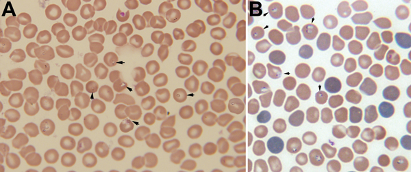
Figure. A) Giemsa-stained thin blood smear for an 8-year-old boy from China showing erythrocytes with typical ring forms, paired pyriforms, and tetrads of a Babesia sp(arrows)B) Giemsa-stained thin blood smear for a mouse with severely combined immunodeficiency, which had been injected with blood from the patient, showing Babesia sp.–infected erythrocytes (arrows)Original magnifications ×1,000.
Page created: April 17, 2014
Page updated: April 17, 2014
Page reviewed: April 17, 2014
The conclusions, findings, and opinions expressed by authors contributing to this journal do not necessarily reflect the official position of the U.S. Department of Health and Human Services, the Public Health Service, the Centers for Disease Control and Prevention, or the authors' affiliated institutions. Use of trade names is for identification only and does not imply endorsement by any of the groups named above.