Volume 21, Number 11—November 2015
Dispatch
Encephalitis-Associated Human Metapneumovirus Pneumonia in Adult, Australia
Anthony Fok12 , Cristina Mateevici, Belinda Lin, Ronil V. Chandra, Victor H.T. Chong1, and Cristina.Mateevici
Figure 2
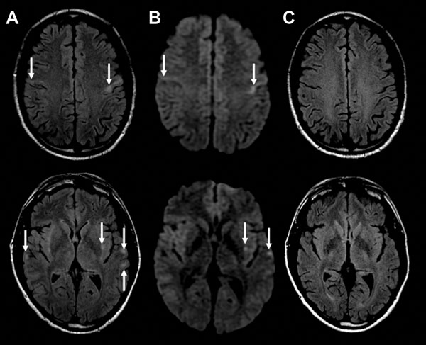
Figure 2. MRI findings from a 47-year-old man with encephalitis-associated human metapneumovirus pneumonia, Australia. A) Axial MRI FLAIR at presentation. Arrows indicate multiple areas of bilateral subcortical and external capsule FLAIR hyperintensities and perirolandic predominance (top image). B) Axial MRI DWI at presentation. Arrows indicate corresponding increase in DWI signal in the affected areas. C) Axial FLAIR MRI after 3 months. The MRI changes have all resolved. DWI, diffusion weighted imaging; FLAIR, fluid-attenuated inversion recovery; MRI, magnetic resonance imaging.
1These authors contributed equally to this article.
2Current affiliation: The University of British Columbia, Vancouver, British Columbia, Canada.
Page created: October 19, 2015
Page updated: October 19, 2015
Page reviewed: October 19, 2015
The conclusions, findings, and opinions expressed by authors contributing to this journal do not necessarily reflect the official position of the U.S. Department of Health and Human Services, the Public Health Service, the Centers for Disease Control and Prevention, or the authors' affiliated institutions. Use of trade names is for identification only and does not imply endorsement by any of the groups named above.