Volume 22, Number 10—October 2016
Dispatch
Reemergence of Mycobacterium chimaera in Heater–Cooler Units despite Intensified Cleaning and Disinfection Protocol
Abstract
Invasive Mycobacterium chimaera infections after open-heart surgery have been reported internationally. These devastating infections result from aerosols generated by contaminated heater–cooler units used with extracorporeal circulation during surgery. Despite intensified cleaning and disinfection, surveillance samples from factory-new units acquired during 2014 grew nontuberculous mycobacteria after a median of 174 days.
Mycobacterium chimaera is an emerging pathogen causing disastrous infections of heart valve prostheses, vascular grafts, and disseminated infections after open-heart surgery (1,2). Growing evidence supports airborne transmission resulting from aerosolization of M. chimaera from contaminated water tanks of heater–cooler units (HCUs) that are used with extracorporeal circulation during surgery (3,4). HCUs were previously associated with surgical site infections caused by nontuberculous mycobacteria (NTM) (5). We describe the colonization dynamics of factory-new HCUs with NTM during regular use.
Identification of M. chimaera infection in 6 patients prompted an outbreak investigation at the University Hospital Zurich, a 900-bed tertiary-care hospital in Zurich, Switzerland, that performs ≈700 open-heart surgeries that use extracorporeal circulation per year. The investigation included microbiologic sampling of HCUs for NTM (3). Surveillance cultures of HCU water from the cardioplegia and patient circuits and airflow samples from running HCUs at ≈2–3 m distance were gathered in monthly intervals. Failure to eradicate M. chimaera and other NTM from older HCUs prompted the acquisition of 5 factory-new HCUs (model 3T; Sorin [now LivaNova, London, UK]) during 2014: 2 in January, 1 in April, and 2 in September.
Mycobacterial cultures were performed according to standard methods by using the mycobacteria growth indicator tube system (MGIT 960; Becton Dickinson, Sparks, MD, USA) or Middlebrook 7H11 agar plates (BD Difco Mycobacteria 7H11 Agar; Becton Dickinson) that were incubated at 37°C for 7 wks or until positive. Air specimens were gathered with a microbiologic air sampler (MAS-100 NT, MBV, Stäfa, Switzerland) running for 2.5 min at a rate of 100 L/min by using Middlebrook 7H11 agar plates. Mycobacterial species were identified by 16S rRNA gene sequencing, as described (6).
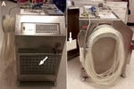
Before mid-April 2014, HCUs were serviced according to the manufacturer’s recommendations. Before first use and every 3 months thereafter, a disinfection cycle was performed by adding 200 mL of 3% sodium hypochlorite (Maranon H; Ecolab, Northwich, UK) to the HCU water tanks filled with filtered tap water (Pall-Aquasafe Water Filter AQ14F1S; Pall, Portsmouth, UK) to a final concentration ≈0.045% sodium hypochlorite in the water tank and circuits. Water was changed every 14 days; 100 mL of 3% hydrogen peroxide was added to the initial water filling (final concentration ≈0.02% hydrogen peroxide in water tank and circuits); an additional 50 mL of 3% hydrogen peroxide was added every 5 days (7). In mid-April 2014, an intensified in-house cleaning and disinfection procedure was implemented, consisting of daily water changes with filtered tap water (Pall) and additions of 100 mL of 3% hydrogen peroxide combined with biweekly disinfection using sodium hypochlorite (Maranon H). In February 2015, with lack of availability of 3% sodium hypochlorite and in line with the manufacturer’s recommendations, the disinfection solution was changed to a combination of peracetic acid and hydrogen peroxide (an additional 450 mL Puristeril 340; Fresenius Medical Care, Hamburg, Germany) in filled water tanks every 2 weeks. Also, stainless steel housings were custom built around the HCUs to ensure strict air separation between the exhaust air of the HCUs and the operating room air (Figure 1). These housings were directly connected to the operating room exhaust conduit.
A total of 134 water samples were obtained from the study HCUs, 127 after implementation of the intensified protocol. The first water sample tested positive for M. chimaera in August 2014, originating from a study HCU introduced 7 months earlier (Figure 2). Of all samples, 90 (67.2%) remained sterile for NTM; 6 (4.5%) were contaminated by bacterial overgrowth; and 38 (28.4%) yielded NTM. Of these NTM, 22 (57.9%) were M. chimaera; 12 (31.6%) were M. gordonae; 1 (2.6%) was M. chelonae; 1 (2.6%) was M. paragordonae; and 2 (5.3%) were a combination of M. chimaera and M. gordonae. NTM were found in both cardioplegia and patient HCU water circuits (Table).
Of 91 air samples, 90 (98.9%) had no mycobacterial growth. One sample grew M. chelonae, although no mycobacterium was detected simultaneously in the corresponding HCU water.
NTM growth was recorded after a median of 174 (range 158–358) days in HCU water samples. One of 5 HCUs remained permanently without growth of M. chimaera; 4 grew M. chimaera after a median of 250 (range 158–358) days.
HCUs seem to provide favorable environmental conditions for growth of NTM, in particular M. chimaera. An intensified cleaning and disinfection protocol failed to prevent growth of NTM entirely but succeeded in preventing detectable aerosolization of M. chimaera.
The contamination status of HCUs seems to be influenced by the intensity of maintenance, especially frequency of water changes. This hypothesis led to the development of the in-house maintenance protocol. The consistently negative air cultures for M. chimaera and the only intermittently positive water cultures support the benefits of our intensified protocol.
Our study design did not elucidate the origin of M. chimaera and other NTM in HCUs. The HCUs might have been already contaminated at time of delivery in a concentration below the detection threshold of mycobacterial cultures. A recent investigation confirmed the presence of environmental mycobacteria, including M. chimaera, in factory-new HCUs (8,9). In our study, 1 HCU (HCU 4, Figure 2) tested positive for M. chimaera for the first time after being returned from repair at the manufacturer. Contamination from tap water is unlikely because the study HCUs used only filtered water.
Previous studies indicated the durability of mycobacteria against several disinfectants, likely because of the organisms’ complex cell wall (10). M. avium complex isolates were shown to have a high level of resistance against chlorine when grown in water (11). Corrosion, certain material characteristics, and dead-end spaces can favor biofilm formation and mycobacterial growth. Killing of NTM with heat may be promising; older studies reported a high efficacy with temperature exposure at 70°C (10,12). A recent report indicated complete suppression of M. chimaera in HCUs by intensified maintenance after a complex decontamination regimen, including replacement of plastic tubing; however, follow-up was limited to 3 months (13). Prolonged testing seems necessary for excluding presence of M. chimaera or other NTM.
Our report has limitations. First, some study HCUs were temporarily maintained according to the manufacturer’s standard before the hospital adopted the intensified protocol. Second, we did not include a control HCU with ongoing maintenance according to the manufacturer’s recommendations. Third, the detection threshold of M. chimaera in water and air cultures remains to be identified. More sensitive culture methods might have produced different results.
Our findings challenge the effectiveness of the HCU manufacturer’s maintenance recommendations, which were recently changed to disinfection with sodium hypochlorite before first use and every 14 days and water changes with all-bacteria–filtered tap water plus 150 mL 3% hydrogen peroxide every 7 days (14). To ensure patient safety until safe HCU technology is available, strict separation of the operating room and HCU air volumes is necessary. This separation can be achieved in several ways. One approach is placing the HCU outside the operating room. Nevertheless, with this measure, the airflow must be restrained from diffusion back into the OR (15). Because the maximum allowed length of water circuit tubing and the architectural layout prohibited this solution at our hospital, we produced airtight housings for the HCUs; however, this solution has less flexible placement of the HCUs within the OR. We continue both the in-house maintenance protocol and regular microbiologic surveillance.
Dr. Schreiber is an internal medicine specialist working as an infectious disease fellow in the Division of Infectious Diseases and Hospital Epidemiology, University Hospital Zurich, Switzerland. His research interests are infections of immunocompromised hosts and infection prevention.
Acknowledgments
We thank the team of perfusionists for obtaining routine HCU water cultures; we also thank Markus Thoma in the Technical Department at University Hospital Zurich for initiating and guiding construction of the custom-built housing for the HCUs.
This study was performed within the framework of the Vascular Graft Cohort Study (VASGRA), which is supported by the Swiss National Science Foundation (grant no. 32473B_163132).
References
- Kohler P, Kuster SP, Bloemberg G, Schulthess B, Frank M, Tanner FC, Healthcare-associated prosthetic heart valve, aortic vascular graft, and disseminated Mycobacterium chimaera infections subsequent to open heart surgery. Eur Heart J. 2015;36:2745–53.DOIPubMedGoogle Scholar
- Centers for Disease Control and Prevention. Non-tuberculous Mycobacterium (NTM) infections and heater-cooler devices interim practical guidance. Updated 2015 Oct 27 [cited 2016 Jun 1]. http://www.cdc.gov/HAI/pdfs/outbreaks/CDC-Notice-Heater-Cooler-Units-final-clean.pdf
- Sax H, Bloemberg G, Hasse B, Sommerstein R, Kohler P, Achermann Y, Prolonged outbreak of Mycobacterium chimaera infection after open-chest heart surgery. Clin Infect Dis. 2015;61:67–75. DOIPubMedGoogle Scholar
- Sommerstein R, Rüegg C, Kohler P, Bloemberg G, Kuster SP, Sax H. Transmission of Mycobacterium chimaera from heater–cooler units during cardiac surgery despite an ultraclean air ventilation system. Emerg Infect Dis. 2016;22:1008–13. DOIPubMedGoogle Scholar
- Nagpal A, Wentink JE, Berbari EF, Aronhalt KC, Wright AJ, Krageschmidt DA, A cluster of Mycobacterium wolinskyi surgical site infections at an academic medical center. Infect Control Hosp Epidemiol. 2014;35:1169–75.DOIPubMedGoogle Scholar
- Bosshard PP, Zbinden R, Abels S, Böddinghaus B, Altwegg M, Böttger EC. 16S rRNA gene sequencing versus the API 20 NE system and the VITEK 2 ID-GNB card for identification of nonfermenting Gram-negative bacteria in the clinical laboratory. J Clin Microbiol. 2006;44:1359–66.DOIPubMedGoogle Scholar
- Sorin Group Deutschland GMBH. Heater-cooler system 3T operating instructions. Version 09/2012. Munich: Sorin Group; 2012.
- Haller S, Höller C, Jacobshagen A, Hamouda O, Abu Sin M, Monnet DL, Contamination during production of heater-cooler units by Mycobacterium chimaera potential cause for invasive cardiovascular infections: results of an outbreak investigation in Germany, April 2015 to February 2016. Euro Surveill. 2016;21:30215. DOIPubMedGoogle Scholar
- US Food and Drug Administration. Mycobacterium chimaera infections associated with Sorin Group Deutschland GmbH Stӧckert 3T Heater-Cooler System: FDA safety communication. 2016 Jun 1 [cited 2016 Jun 1]. http://www.fda.gov/MedicalDevices/Safety/AlertsandNotices/ucm504213.htm
- Vaerewijck MJ, Huys G, Palomino JC, Swings J, Portaels F. Mycobacteria in drinking water distribution systems: ecology and significance for human health. FEMS Microbiol Rev. 2005;29:911–34.DOIPubMedGoogle Scholar
- Taylor RH, Falkinham JO III, Norton CD, LeChevallier MW. Chlorine, chloramine, chlorine dioxide, and ozone susceptibility of Mycobacterium avium. Appl Environ Microbiol. 2000;66:1702–5.DOIPubMedGoogle Scholar
- Schulze-Röbbecke R, Buchholtz K. Heat susceptibility of aquatic mycobacteria. Appl Environ Microbiol. 1992;58:1869–73.PubMedGoogle Scholar
- Garvey MI, Ashford R, Bradley CW, Bradley CR, Martin TA, Walker J, Decontamination of heater-cooler units associated with contamination by atypical mycobacteria. J Hosp Infect. 2016;93:229–34.DOIPubMedGoogle Scholar
- Sorin Group Deutschland GMBH. Heater-cooler system 3T operating instructions. 2015 Feb [cited 2016 Jun 1]. http://www.sorineifu.com/PDFs/45-91-45USA_C.PDF
- Götting T, Klassen S, Jonas D, Benk C, Serr A, Wagner D, Heater-cooler units: contamination of crucial devices in cardiothoracic surgery. J Hosp Infect. 2016;93:223–8.DOIPubMedGoogle Scholar
Figures
Table
Cite This Article1Current affiliation: Unilabs, Dübendorf, Switzerland.
Table of Contents – Volume 22, Number 10—October 2016
| EID Search Options |
|---|
|
|
|
|
|
|


Please use the form below to submit correspondence to the authors or contact them at the following address:
Hugo Sax, Division of Infectious Diseases and Hospital Epidemiology, University Hospital and University of Zurich, Rämistrasse 100, CH-8091 Zurich, Switzerland
Top