Skip directly to site content Skip directly to page options Skip directly to A-Z link Skip directly to A-Z link Skip directly to A-Z link
Volume 22, Number 11—November 2016
Research

Ambulatory Pediatric Surveillance of Hand, Foot and Mouth Disease as Signal of an Outbreak of Coxsackievirus A6 Infections, France, 2014–2015

Audrey MirandComments to Author , François Vié le Sage, Bruno Pereira, Robert Cohen, Corinne Levy, Christine Archimbaud, Hélène Peigue-Lafeuille, Jean-Luc Bailly, and Cécile Henquell
Author affiliations: Centre Hospitalier Universitaire de Clermont-Ferrand, Clermont-Ferrand, France (A. Mirand, B. Pereira, C. Archimbaud, H. Lafeuille, J.-L. Bailly, C. Henquell); Université d’Auvergne, Clermont-Ferrand (A. Mirand, C. Archimbaud, H. Peigue-Lafeuille, J.-L. Bailly, C. Henquell); Association Française de Pédiatrie Ambulatoire, Bagnols-sur-Cèze, France (F. Vié le Sage); Association Clinique et Thérapeutique Infantile du Val de Marne, Saint Maur des Fossés, France (R. Cohen, C. Levy); Université Paris Est, Créteil, France (R. Cohen, C. Levy)

Main Article

Figure 4

Phylogenetic tree based on partial viral protein (VP1) coding sequences of coxsackievirus (CV) A6, France, April 2014–March 2015. The maximum credibility tree is inferred with the partial VP1 sequence (369 nt, position 2,441–2,808 relative to the Gdula CV-A6 prototype strain). The phylogenetic relationships were inferred with a Bayesian method by using a relaxed molecular clock model. The tree was reconstructed using Figtree version 1.4.2 (http://tree.bio.ed.ac.uk/software/figtree). For clarity,

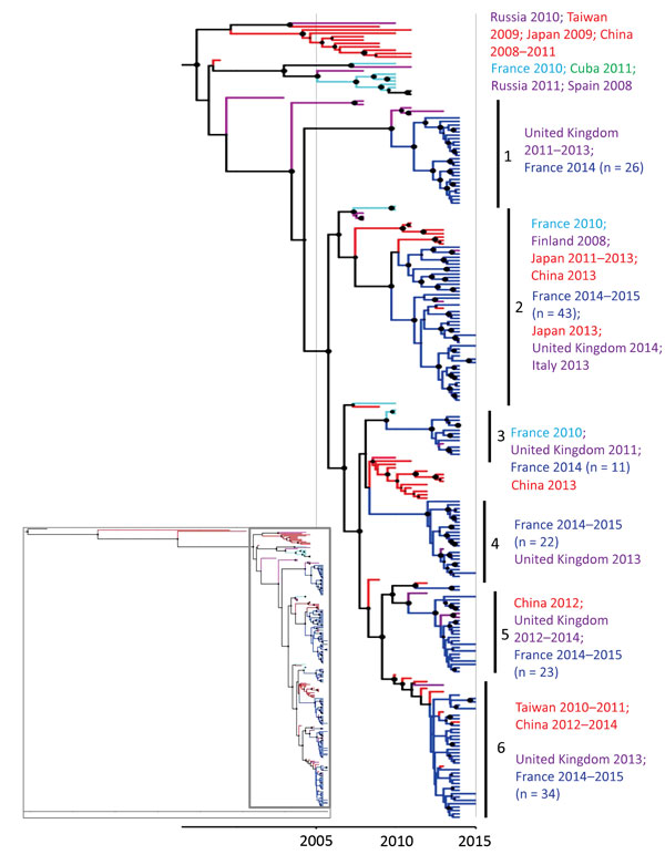
Figure 4. Phylogenetic tree based on partial viral protein (VP1) coding sequences of coxsackievirus (CV) A6, France, April 2014–March 2015. The maximum credibility tree is inferred with the partial VP1 sequence (369 nt, position 2,441–2,808 relative to the Gdula CV-A6 prototype strain). The phylogenetic relationships were inferred with a Bayesian method by using a relaxed molecular clock model. The tree was reconstructed using Figtree version 1.4.2 (http://tree.bio.ed.ac.uk/software/figtree). For clarity, the sequence names are not included in the tree. Circle sizes are proportional to posterior probability. Each tip branch represents a sampled virus sequence. The continents/countries where the virus strains were sampled are indicated by different colors: Europe, purple; France 2010, light blue; France 2014–2015, dark blue; the Americas, green; Asia, red. The inset shows the complete tree, with the box indicating the portion enlarged for clarity.

Main Article

Page created: October 18, 2016
Page updated: October 18, 2016
Page reviewed: October 18, 2016
The conclusions, findings, and opinions expressed by authors contributing to this journal do not necessarily reflect the official position of the U.S. Department of Health and Human Services, the Public Health Service, the Centers for Disease Control and Prevention, or the authors' affiliated institutions. Use of trade names is for identification only and does not imply endorsement by any of the groups named above.
file_external