Volume 22, Number 12—December 2016
Dispatch
Human Infection with Novel Spotted Fever Group Rickettsia Genotype, China, 2015
Cite This Article
Citation for Media
Abstract
Only 4 species of spotted fever group rickettsiae have been detected in humans in China. However, phylogenetic analysis of samples from 5 ill patients in China indicated infection with a novel spotted fever group Rickettsia, designated Rickettsia sp. XY99. Clinical signs resembled those of severe fever with thrombocytopenia syndrome.
Spotted fever group (SFG) rickettsiae are globally distributed and mostly transmitted by ticks (1). Recently, emerging and reemerging SFG rickettsiae, such as Rickettsia slovaca (2), R. aeschlimannii (3), R. massiliae (4), Candidatus Rickettsia tarasevichiae (5,6), and R. sibirica subspecies sibirica BJ-90 (7), previously considered nonpathogenic, were found to infect humans. In addition, R. parkeri was confirmed to be pathogenic 65 years after its detection in ticks in 1939 (8).
In China, SFG rickettsioses are not listed as reportable diseases, and only 4 species of SFG rickettsiae (R. heilongjiangensis, R. sibirica subspecies sibirica BJ-90, Candidatus Rickettsia tarasevichiae, and R. raoultii) have been detected in human blood samples (9). In contrast, besides these pathogenic species, at least 4 other species of SFG rickettsiae (R. sibirica subspecies mongolotimonae, R. monacensis, R. slovaca, Candidatus Rickettsia hebeiii) have been detected in ticks, urging a wider search for cases in humans. We report infection of 5 patients with a novel SFG rickettsia in eastern central China.
From March through November 2015, at the People’s Liberation Army 154 Hospital in Xinyang City, Henan Province, China, patients who were acutely symptomatic with fever and had a history of tick bites or animal contact within the past month were screened for SFG rickettsiae infection. At admission, EDTA-anticoagulated samples of peripheral blood were collected. DNA was extracted by using a QIAamp DNA Blood Mini Kit (QIAGEN, Germantown, MD, USA). Nested PCRs selective for outer membrane protein A (ompA) and citrate synthase (gltA) genes were concurrently performed to detect SFG rickettsial DNA (Technical Appendix Table 1). Positive amplicons were purified and then sequenced in both directions. Acute-phase (<7 days after illness onset) and convalescent-phase (>14 days after illness onset) serum samples were tested by indirect immunofluorescence assay (IFA) for IgG against R. rickettsii by using a commercially available IFA kit (Focus Diagnostics Inc., Cypress, CA, USA).
Figure 1
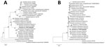
Figure 1. Phylogenetic analyses based on nucleotide sequences of the outer member protein A (307-bp) (A) and citrate synthase (1,150-bp) (B) genes of Rickettsia. Boldface indicates the newly discovered Rickettsia genotype (Rickettsia sp....
Positive amplification of ompA and gltA genes was found for 5 patients, and the obtained sequences for each of the 2 genes from all 5 patients were identical. Nucleotide sequence (350-bp) of ompA gene (GenBank accession no. KU853020) from each of the 5 patients showed 10-bp differences from that of R. massiliae strain AZT80 (GenBank accession no. CP003319) and 12-bp differences from that of R. rhipicephali strain HJ#5 (GenBank accession no. CP013133). Nucleotide sequences (1150-bp) of gltA gene (GenBank accession no. KU853022) from each of the 5 patients differed from that of R. massiliae strain AZT80 by 4 bp and from that of R. rhipicephali strain HJ#5 by 5 bp (Technical Appendix Table 2). According to phylogenetic analysis, the novel SFG rickettsiae genotype, here designated as Rickettsia sp. XY99, seems to represent a distinct lineage and could constitute a new species (Figure 1). For all 5 patients, seroconversion or a 4-fold increase of IgG against R. rickettsii was found between the acute- and convalescent-phase samples, and the patients were determined to have acute infection with SFG rickettsiae (Technical Appendix Table 3). Subsequent testing of the 5 patients for infection with severe fever with thrombocytopenia syndrome virus, Anaplasma phagocytophilum, “A. capra,” and Babesia microti by molecular (real-time PCR or nested PCR) and serologic tests (ELISA or IFA) produced no positive results.
Figure 2
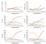
Figure 2. Dynamic changes of 6 laboratory parameters (with 2-day intervals) during hospitalization of 5 patients with Rickettsia sp. XY99 infection, China, 2015. Red, patient 1; yellow, patient 2; green, patient 3; purple,...
All 5 patients were farmers who resided in the villages of Xinyang City. Patient median age was 65 (range 62–80) years, and 3 were male (Table). Two patients had a history of tick exposure, and the other 3 had had contact with livestock. For all 5 patients, illness onset occurred June 20–July 10, 2015. The median time from illness onset to admission was 4 (range 3–6) days, and the median duration of hospitalization was 10 (range 8–12) days. All patients experienced fever (highest 38.4°C– 40.0°C), asthenia, anorexia, and nausea; 4 had cough, 3 vomiting, 2 myalgia, 1 headache, and 1 dizziness. Of note, all 5 patients had lymphadenopathy, but none had rash or eschar. At admission, all 5 patients had leukopenia, thrombocytopenia, and elevated hepatic aminotransferase levels; 4 had elevated lactate dehydrogenase levels, and 2 had elevated creatine kinase levels (Figure 2). Treatment included therapy with cefminox and cefoperazone; no doxycycline was used.
Complications included pneumonia (3 patients), hemorrhagic signs (3), hydrothorax (2), and neurologic syndromes (1). For 1 patient, severe complications progressively emerged 6 days after disease onset and included pneumonia and hydrothorax (Technical Appendix Figure), confusion, meningeal irritation sign, ecchymosis, and hematuria. Laboratory indicators were substantially more out of range 7 days after disease onset, indicative of severe multiorgan dysfunction (Figure 2). Treatment was ineffective, and the patient died 15 days after disease onset. The other 4 patients were discharged after 8–12 days’ hospitalization; at that time, all clinical signs and symptoms had resolved, but for certain patients, laboratory values remained out of reference range, suggesting slow recovery of organ dysfunction (Figure 2).
Our detection of Rickettsia sp. XY99 DNA in blood samples collected during the acute period of illness (days 3–6 after onset) from 5 patients in the same region of China suggests that this organism was the etiologic agent of the infection. Seroconversion or a 4-fold increase in titers of IgG against R. rickettsii provided supportive evidence of SFG Rickettsia infection. Phylogenetic analysis indicated that Rickettsia sp. XY99 was a novel genotype of SFG rickettsiae.
In contrast to humans with R. massiliae infection and many other SFG rickettsioses reported previously (4,10), none of the 5 patients infected with Rickettsia sp. XY99 had rash or eschar, and only 1 had headache. In recent years, the concept of the classic triad of fever, rash, and headache suggesting infection with SFG rickettsiae has been increasingly challenged. Several emerging SFG rickettsiae species, such as R. slovaca (2), R. raoultii (11), R. africae (12), and R. helvetica (13), can infect humans, but such infections lack these traditional features, which were also lacking in the cases reported here. Therefore, absence of rash and tick-bite history should not exclude suspicion of SFG rickettsiae infection.
Similar to R. slovaca and R. raoultii infections, which can be associated with tickborne lymphadenopathy and Dermacentor-borne necrosis-erythema-lymphadenopathy (14), lymphadenopathy was also observed in all 5 patients with Rickettsia sp. XY99 infection. Thus, lymphadenopathy might be a typical sign useful for clinical diagnosis of Rickettsia sp. XY99 infection. All 5 patients had gastrointestinal syndromes, indicating potential tissue lesions or vascular injury of the gastrointestinal tract. The hydrothorax and multiple hemorrhagic signs in 4 patients is possibly suggestive of vascular invasion or damage caused by this novel Rickettsia species.
Confirmation of the novel Rickettsia genotype was achieved only by sequencing the ompA and gltA genes. Although differences based on 2 gene segments support its designation as a novel species, isolation efforts and characterization of all 5 genes (rrs, gltA, ompA, ompB, and geneD) are warranted, according to the guidelines for classification of a new Rickettsia species (15).
Physicians in this area of China should be aware of human infections with Rickettsia sp. XY99. It should be included in differential diagnoses with severe fever with thrombocytopenia syndrome, which causes similar clinical illness and is also endemic to the same area in eastern central China.
Dr. Li is a scientist in the State Key Laboratory of Pathogen and Biosecurity, Beijing Institute of Microbiology and Epidemiology. His research interests include microbiology, epidemiology, and ecology of tickborne diseases.
Acknowledgment
This study was supported by the Natural Science Foundation of China (81222037, 81290344, 81130086, and 81472005).
References
- Parola P, Paddock CD, Socolovschi C, Labruna MB, Mediannikov O, Kernif T, et al. Update on tick-borne rickettsioses around the world: a geographic approach. Clin Microbiol Rev. 2013;26:657–702.DOIPubMedGoogle Scholar
- Raoult D, Berbis P, Roux V, Xu W, Maurin M. A new tick-transmitted disease due to Rickettsia slovaca. Lancet. 1997;350:112–3.DOIPubMedGoogle Scholar
- Raoult D, Fournier PE, Abboud P, Caron F. First documented human Rickettsia aeschlimannii infection. Emerg Infect Dis. 2002;8:748–9.DOIPubMedGoogle Scholar
- Vitale G, Mansuelo S, Rolain JM, Raoult D. Rickettsia massiliae human isolation. Emerg Infect Dis. 2006;12:174–5.DOIPubMedGoogle Scholar
- Jia N, Zheng YC, Jiang JF, Ma L, Cao WC. Human infection with Candidatus Rickettsia tarasevichiae. N Engl J Med. 2013;369:1178–80.DOIPubMedGoogle Scholar
- Liu W, Li H, Lu QB, Cui N, Yang ZD, Hu JG, et al. Candidatus Rickettsia tarasevichiae infection in eastern central China: a case series. Ann Intern Med. 2016;164:641–8.DOIPubMedGoogle Scholar
- Jia N, Jiang JF, Huo QB, Jiang BG, Cao WC. Rickettsia sibirica subspecies sibirica BJ-90 as a cause of human disease. N Engl J Med. 2013;369:1176–8.DOIPubMedGoogle Scholar
- Paddock CD, Sumner JW, Comer JA, Zaki SR, Goldsmith CS, Goddard J, et al. Rickettsia parkeri: a newly recognized cause of spotted fever rickettsiosis in the United States. Clin Infect Dis. 2004;38:805–11.DOIPubMedGoogle Scholar
- Fang LQ, Liu K, Li XL, Liang S, Yang Y, Yao HW, et al. Emerging tick-borne infections in mainland China: an increasing public health threat. Lancet Infect Dis. 2015;15:1467–79.DOIPubMedGoogle Scholar
- Cascio A, Torina A, Valenzise M, Blanda V, Camarda N, Bombaci S, et al. Scalp eschar and neck lymphadenopathy caused by Rickettsia massiliae. Emerg Infect Dis. 2013;19:836–7.DOIPubMedGoogle Scholar
- Parola P, Rovery C, Rolain JM, Brouqui P, Davoust B, Raoult D. Rickettsia slovaca and R. raoultii in tick-borne Rickettsioses. Emerg Infect Dis. 2009;15:1105–8.DOIPubMedGoogle Scholar
- Raoult D, Fournier PE, Fenollar F, Jensenius M, Prioe T, de Pina JJ, et al. Rickettsia africae, a tick-borne pathogen in travelers to sub-Saharan Africa. N Engl J Med. 2001;344:1504–10.DOIPubMedGoogle Scholar
- Fournier PE, Allombert C, Supputamongkol Y, Caruso G, Brouqui P, Raoult D. Aneruptive fever associated with antibodies to Rickettsia helvetica in Europe and Thailand. J Clin Microbiol. 2004;42:816–8.DOIPubMedGoogle Scholar
- Silva-Pinto A, Santos ML, Sarmento A. Tick-borne lymphadenopathy, an emerging disease. Ticks Tick Borne Dis. 2014;5:656–9.DOIPubMedGoogle Scholar
- Fournier PE, Dumler JS, Greub G, Zhang J, Wu Y, Raoult D. Gene sequence-based criteria for identification of new rickettsia isolates and description of Rickettsia heilongjiangensis sp. nov. J Clin Microbiol. 2003;41:5456–65.DOIPubMedGoogle Scholar
Figures
Table
Cite This Article1These authors contributed equally to this article.
Table of Contents – Volume 22, Number 12—December 2016
| EID Search Options |
|---|
|
|
|
|
|
|
Please use the form below to submit correspondence to the authors or contact them at the following address:
Address for correspondence: Wu-Chun Cao or Wei Liu, State Key Laboratory of Pathogen and Biosecurity, Beijing Institute of Microbiology and Epidemiology, 20 Dong-Da St, Fengtai District, Beijing 100071, China; or
Top