Volume 22, Number 6—June 2016
Letter
MERS-CoV Infection of Alpaca in a Region Where MERS-CoV is Endemic
Figure
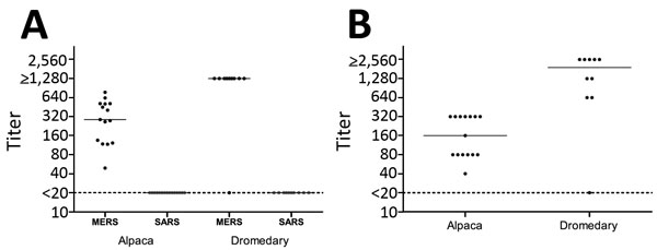
Figure. Column scatterplots of MERS-CoV reactivity of serum samples from alpaca (n = 15) and dromedaries (n = 10) in the Al Shahaniya region of Qatar, April 2015. A) Plot of alpaca and dromedary serum titers of antibodies specific for S1 antigens of 2 coronaviruses as determined by protein microarray. Titers were defined as the interpolated serum concentration that provoked a response half-way on a concentration-response curve between the minimum and maximum signal and were calculated from the inflection point of a 4-step dilution series (1:20 to 1:1,280) as described previously (5). B) Plot of alpaca and dromedary serum titers of MERS-CoV neutralizing antibodies as determined by PRNT90. The highest serum dilution neutralizing 90% of plaque formation is depicted. For both panels, solid lines indicate median, and dotted lines indicate detection limit. MERS, Middle East respiratory syndrome; CoV, coronavirus; PRNT90, 90% plaque-reduction neutralization test; SARS, severe acute respiratory syndrome.
References
- Chan JF, Lau SK, To KK, Cheng VC, Woo PC, Yuen KY. Middle East respiratory syndrome coronavirus: another zoonotic betacoronavirus causing SARS-like disease. Clin Microbiol Rev. 2015;28:465–522. DOIPubMedGoogle Scholar
- Haagmans BL, Al Dhahiry SH, Reusken CB, Raj VS, Galiano M, Myers R, Middle East respiratory syndrome coronavirus in dromedary camels: an outbreak investigation. Lancet Infect Dis. 2014;14:140–5.PubMedGoogle Scholar
- Reusken CB, Farag EA, Haagmans BL, Mohran KA, Godeke GJVV, Raj S, Occupational exposure to dromedaries and risk for MERS-CoV infection, Qatar, 2013–2014. Emerg Infect Dis. 2015;21:1422–5. DOIPubMedGoogle Scholar
- Reusken CB, Haagmans BL, Müller MA, Gutierrez C, Godeke GJ, Meyer B, Middle East respiratory syndrome coronavirus neutralising serum antibodies in dromedary camels: a comparative serological study. Lancet Infect Dis. 2013;13:859–66. DOIPubMedGoogle Scholar
- Koopmans M, de Bruin E, Godeke GJ, Friesema I, van Gageldonk R, Schipper M, Profiling of humoral immune responses to influenza viruses by using protein microarray. Clin Microbiol Infect. 2012;18:797–807. DOIPubMedGoogle Scholar
- Corman VM, Eckerle I, Bleicker T, Zaki A, Landt O, Eschbach-Bludau M, Detection of a novel human coronavirus by real-time reverse-transcription polymerase chain reaction. Euro Surveill. 2012;17:20285 .PubMedGoogle Scholar
- Sabir JS, Lam TT, Ahmed MM, Li L, Shen Y, Abo-Aba SE, Co-circulation of three camel coronavirus species and recombination of MERS-CoVs in Saudi Arabia. Science. 2016;351:81–4. DOIPubMedGoogle Scholar
- Eckerle I, Corman VM, Müller MA, Lenk M, Ulrich RG, Drosten C. Replicative capacity of MERS coronavirus in livestock cell lines. Emerg Infect Dis. 2014;20:276–9 . DOIPubMedGoogle Scholar
- Chan SM, Damdinjav B, Perera RA, Chu DK, Khishgee B, Enkhbold B, Absence of MERS-coronavirus in Bactrian camels, southern Mongolia, November 2014. Emerg Infect Dis. 2015;21:1269–71. DOIPubMedGoogle Scholar
- Wernery U, Kinne J. Foot and mouth disease and similar virus infections in camelids: a review. Rev Sci Tech. 2012;31:907–18 .PubMedGoogle Scholar
1These authors contributed equally to this article.