Volume 22, Number 7—July 2016
Letter
Senecavirus A in Pigs, United States, 2015
Ben Hause , Olivia Myers, Joshua Duff, and Richard A. Hesse
Figure
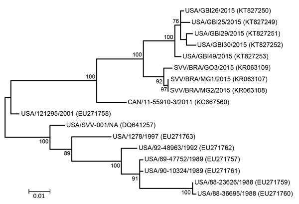
Figure. Phylogenetic tree of Senecavirus A P1 sequences. Maximum-likelihood analysis in combination with 1,000 bootstrap replicates as implemented in MEGA 6.06 (http://www.megasoftware.net) was used to derive the tree on the basis of nucleotide sequences. GenBank accession numbers are shown in parentheses. SVV in some isolate names indicates Seneca Valley virus, the original name for Senecavirus A. Scale bar indicates number of nucleotide changes per site.
Page created: June 14, 2016
Page updated: June 14, 2016
Page reviewed: June 14, 2016
The conclusions, findings, and opinions expressed by authors contributing to this journal do not necessarily reflect the official position of the U.S. Department of Health and Human Services, the Public Health Service, the Centers for Disease Control and Prevention, or the authors' affiliated institutions. Use of trade names is for identification only and does not imply endorsement by any of the groups named above.