Volume 22, Number 7—July 2016
Letter
Crimean-Congo Hemorrhagic Fever with Acute Subdural Hematoma, Mauritania, 2012
Ahmed S. Kleib , Sidi M. Salihy, Sidi M. Ghaber, Baba W. Sidiel, Khalil C. Sidiya, and Ely S. Bettar1
Figure
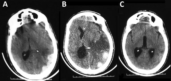
Figure. Computed tomography scan image of the brain of a 58-year-old man with Crimean-Congo hemorrhagic fever, Mauritania, 2012. A) Acute subdural hematoma, on the left side. B) Subdural hematoma with perihematomal edema and midline shift. C) Complete resorption of the subdural hematoma with residual edema, 1 month later.
1All authors contributed equally to this article.
Page created: June 14, 2016
Page updated: June 14, 2016
Page reviewed: June 14, 2016
The conclusions, findings, and opinions expressed by authors contributing to this journal do not necessarily reflect the official position of the U.S. Department of Health and Human Services, the Public Health Service, the Centers for Disease Control and Prevention, or the authors' affiliated institutions. Use of trade names is for identification only and does not imply endorsement by any of the groups named above.