Skip directly to site content Skip directly to page options Skip directly to A-Z link Skip directly to A-Z link Skip directly to A-Z link
Volume 23, Number 11—November 2017
Research

Airborne Transmission of Highly Pathogenic Influenza Virus during Processing of Infected Poultry

Kateri Bertran, Charles Balzli, Yong-Kuk Kwon1, Terrence M. Tumpey, Andrew Clark, and David E. SwayneComments to Author 
Author affiliations: US Department of Agriculture, Athens, Georgia, USA (K. Bertran, C. Balzli, Y.-K. Kwon, D.E. Swayne); Centers for Disease Control and Prevention, Atlanta, Georgia, USA (T.M. Tumpey); International Veterinary Consultant, Pendleton, Oregon, USA (A. Clark)

Main Article

Figure 3

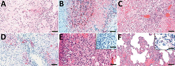
Histologic lesions and immunohistochemical detection of viral antigen in ferrets exposed to LPM processing of A/Vietnam/1203/04(H5N1) HPAI virus–infected chickens. A) Olfactory bulb, 7 dpe. Diffuse and severe neuropil malacia with mild cavitation and focal hemorrhages. Scale bar = 50 μm. B) Olfactory bulb, 7 dpe. Viral antigen detected in neuropil, astrocytes, and neurons. Scale bar = 50 μm. C) Liver, 8 dpe. Confluent coagulative necrosis of hepatocytes and bile duct necrosis with mononuclear ce

Figure 3. Histologic lesions and immunohistochemical detection of viral antigen in samples from ferrets exposed to live poultry market processing of highly pathogenic avian influenza A/Vietnam/1203/04 (H5N1) virus–infected chickens in study of airborne transmission of highly pathogenic influenza virus during processing of infected poultry. A) Olfactory bulb, 7 dpe, showing diffuse and severe neuropil malacia with mild cavitation and focal hemorrhages. Scale bar = 50 μm. B) Olfactory bulb, 7 dpe, showing viral antigen detected in neuropil, astrocytes, and neurons. Scale bar = 50 μm. C) Liver, 8 dpe, showing confluent coagulative necrosis of hepatocytes and bile duct necrosis with mononuclear cellular infiltrate in the portal triad. Scale bar = 50 μm. D) Liver, 8 dpe, showing viral antigen detected in hepatocytes, bile duct epithelia, and cellular debris. Scale bar = 50 μm. E) Nasal cavity, 7 dpe, showing moderate necrotic rhinitis with coagulative necrosis of mucous glandular epithelial cells; insert shows no viral antigen detected in mucosal membrane. Scale bars = 25 μm. F) Lung, 7 dpe, showing mild histiocytic interstitial pneumonia; insert shows viral antigen detected in type II pneumocytes. Scale bars = 25 μm. dpe, days postexposure.

Main Article

1Current affiliation: Animal and Plant Quarantine Agency, Gimcheon-si, South Korea.

Page created: October 26, 2017
Page updated: October 26, 2017
Page reviewed: October 26, 2017
The conclusions, findings, and opinions expressed by authors contributing to this journal do not necessarily reflect the official position of the U.S. Department of Health and Human Services, the Public Health Service, the Centers for Disease Control and Prevention, or the authors' affiliated institutions. Use of trade names is for identification only and does not imply endorsement by any of the groups named above.
file_external