Volume 23, Number 12—December 2017
Research
Evolutionary Context of Non–Sorbitol-Fermenting Shiga Toxin–Producing Escherichia coli O55:H7
Cite This Article
Citation for Media
Abstract
In July 2014, an outbreak of Shiga toxin–producing Escherichia coli (STEC) O55:H7 in England involved 31 patients, 13 (42%) of whom had hemolytic uremic syndrome. Isolates were sequenced, and the sequences were compared with publicly available sequences of E. coli O55:H7 and O157:H7. A core-genome phylogeny of the evolutionary history of the STEC O55:H7 outbreak strain revealed that the most parsimonious model was a progenitor enteropathogenic O55:H7 sorbitol-fermenting strain, lysogenized by a Shiga toxin (Stx) 2a–encoding phage, followed by loss of the ability to ferment sorbitol because of a non-sense mutation in srlA. The parallel, convergent evolutionary histories of STEC O157:H7 and STEC O55:H7 may indicate a common driver in the evolutionary process. Because emergence of STEC O157:H7 as a clinically significant pathogen was associated with acquisition of the Stx2a-encoding phage, the emergence of STEC O55:H7 harboring the stx2a gene is of public health concern.
The first outbreak of Shiga–toxin producing Escherichia coli (STEC) O55:H7 in the United Kingdom occurred in the county of Dorset, England, in July 2014 (1). Ultimately, 31 cases were linked to the outbreak, and 13 (42%) of those patients had hemolytic uremic syndrome (HUS). Of the 13 with HUS, 8 (66%) had neurologic complications and 11 (90%) required prolonged treatment for kidney replacement. After enhanced epidemiologic surveillance and analysis of the patients’ food, exposure, and travel histories, the only epidemiologic link identified was living in or having close links to Dorset County. Extensive microbiological investigations included testing of the environment, nondomestic animals, and household pets. Although no causal link was established, whole-genome sequencing and epidemiologic analyses were indicative of a local endemic zoonotic source (1).
Previous studies postulated that the common STEC O157:H7 clone evolved from enteropathogenic E. coli (EPEC) serotype O55:H7 (2,3). Evolutionary models predict the stepwise acquisition of a Shiga toxin (Stx)–encoding bacteriophage in the EPEC O55:H7 progenitor strain, followed by the substitution of the rfb locus encoding the somatic O55 antigen with that encoding the O157 antigen, the acquisition of the pO157 plasmid, loss of the ability to ferment sorbitol, and loss of the ability to produce β-glucuronidase (3–6). Analyses from more recent studies have indicated that the Stx-encoding phage is an unstable evolutionary marker, with frequent acquisition and loss occurring in STEC O55:H7 and all 3 lineages of STEC O157 throughout their evolutionary history (7,8).
STEC O157:H7 has multiple genetic and phenotypic features that contribute to its pathogenicity or are used for detection and identification. The primary virulence factor defining the STEC group is production of Stx1, Stx2, or both. The genes encoding the toxins, stx1 and stx2, are harbored on lambdoid prophage and are the targets of commercial and in-house diagnostic PCR assays (9). Both toxins can be divided into several subtypes, Stx1a–1d and Stx2a–2g (10). The locus of enterocyte effacement (LEE) is a 35-kb pathogenicity island encoding a type III secretion system (T3SS) responsible for the attaching and effacing phenotype that facilitates successful colonization of the human gut (11). The inability to ferment sorbitol or to produce β-glucuronidase differentiates STEC O157 from ≈90% of other gastrointestinal bacteria (5,12). These characteristics, along with resistance to tellurite, facilitate the detection and identification of STEC O157:H7 on selective media. The pO157 plasmid encodes multiple putative virulence factors, including enterohemolysin (ehxA) and an adhesin (toxB) (13).
The STEC O55:H7 Dorset outbreak strain shared certain characteristics with the STEC O157:H7 clone. Initial PCRs detected the presence of stx2 and the intimin gene eae, a marker for E. coli attaching and effacing phenotype; non–sorbitol-fermenting colonies of STEC O55 were identified after culture on sorbitol MacConkey agar (1,9). However, unlike the STEC O157 clone, the STEC O55 Dorset outbreak strain exhibited β-glucuronidase activity and was sensitive to tellurite. Laboratory records held at the Gastrointestinal Bacterial Reference Unit of Public Health England showed that this highly pathogenic strain had not previously been isolated from humans or animals in the United Kingdom. Our goal with this study was to identify the genetic determinants responsible for the phenotypic characteristics of the STEC O55:H7 Dorset outbreak strain and to explore the strain’s evolutionary history.
Bacterial Strains
We studied 26 isolates of STEC O55:H7 from the outbreak, 10 isolates of STEC O55:H7 from Ireland, and 79 isolates selected to represent of the broad phylogeny of STEC O157:H7 (Technical Appendix Table). From public databases, we retrieved 10 genome sequences for E. coli O55:H7 and 2 for STEC O157:H7 (6,7,14,15) (Table 1).
Whole-Genome Sequencing, Assembly, and Alignment
We sequenced all isolates by using an Illumina paired-end (100-bp) protocol (https://www.illumina.com) and assembled them by using SPAdes Genome Assembler version 3.1.1 (18). The assemblies were annotated by using Prokka version 1.0.1 (19). We used the MinION (https://nanoporetech.com/products/minion) nanopore platform to sequence an isolate from the outbreak, designated 122262. A hybrid Illumina/MinION de novo assembly of 122262 constructed by using SPAdes yielded 15 contigs with the largest contig spanning the first 2.4 mbp. We aligned published reference genomes against the outbreak reference strain 122262 by using Mauve (20).
Genome, Plasmid, and Bacteriophage Comparisons
We retrieved from GenBank published nucleotide sequences of key virulence genes associated with toxicity, host-cell adhesion, and metabolic activity and concatenated in FASTA (http://www.ebi.ac.uk/Tools/sss/fasta/) file format. To determine the presence and absence of the gene panel, we performed a blastn (21) comparison against the extracted coding sequences of 122262. Significant hits were defined as those with a nucleotide identity of >90% over at least 90% of the query sequence. Truncated sequences were defined as matches with <90% coverage. We uploaded assembled data from the strains in FASTA file format to the PHAge Search Tool (PHAST) web server for prophage identification (22). Prophage region detection, prophage annotation, and circular genomic views from PHAST results were used along blast ring image generator (BRIG) plots (23) to isolate the prophage regions of 122262 and nucleotide homologies to the prophages in the Sakai reference genome (16). BRIG was used to visually compare the similarities between the Sakai and outbreak strain prophages. We compared prophage regions of 122262 with those extracted and analyzed by Shaaban et al. (17) by using the pipeline and strains presented in their study.
Phylogenetic Analyses
Short reads were quality trimmed (24) and mapped to the STEC O157:H7 Sakai reference genome (GenBank accession no. BA000007) by using Burrows-Wheeler aligner–maximal exact matching (25). We sorted and indexed the sequence alignment map output from the Burrows-Wheeler aligner to produce a binary alignment map by using SAMtools (25). GATK2 (26) was used to create a variant call format file from each of the B binary alignment maps, which were further parsed to extract only single-nucleotide polymorphism (SNP) positions that were of high quality (mapping quality >30, coverage of reads that passed quality metrics >10, variant ratio >0.9). We used pseudosequences of polymorphic positions to create maximum-likelihood trees by using RAxML (27). FASTQ (https://www.ncbi.nlm.nih.gov/pmc/articles/PMC2847217/) sequences were deposited in the National Center for Biotechnology Information Short Read Archive under the BioProject PRJNA248042.
General Genomic Features
STEC O55:H7 strain 122262 had a 5,364,131-bp chromosome and a 67,247-bp single plasmid of replicon type FIB-15. Use of blastn to compare the extracted plasmid sequence from 122262 with publicly available plasmid sequences belonging to CB9615, 2013C-4465, and Sakai indicated that the plasmid of 122262 was 99% identical to pO55 CB9615 over its complete length. Unlike pO157 in STEC O157:H7, the O55:H7 plasmids did not encode toxin B (toxB) or the enterohemolysin operon (ehxABCD). The E. coli O55:H7 strains 122262, CB9615, and 2013C-4465 did, however, encode a remote toxB homologue efa1/lifA on the chromosome that has 29% nt identity (97% coverage) with pO157 toxB. The LEE was inserted into the chromosome of strain 122262 at tRNA-selC, the most common insertion site in a range of pathogenic E. coli chromosomal backgrounds (28). Antimicrobial drug resistance determinants included aadA-1b encoding resistance to streptomycin and dfrA-1 encoding resistance to trimethoprim.
Prophage Composition of 122262
Figure 1

PHAST identified 15 prophage interruptions in 122262, of which 5 were homologous in nucleotide identity to Sp2, Sp3, Sp6, Sp8, and Sp14 found in Sakai (16) (Table 2; Figure 1). Unique genetic content and position was found for 9 putative prophages (Figure 1). In addition, a Stx2a-encoding phage was identified at the Stx-associated bacteriophage insertion site yecE in strain 122262. In Sakai, the Stx1a (Sp15) and Stx2a (Sp5) encoding phages are inserted at wrbA and yehV, respectively. However, yecE is a known Stx-associated bacteriophage insertion site in strains of STEC O157:H7 encoding stx2a belonging to lineage Ic (29).
Figure 2
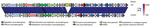
Figure 2. The sequence of the Stx2a-encoding phage from the July 2014 Dorset County, England, outbreak strain of Shiga toxin–producing Escherichia coli O55:H7, designated 122262, showed >98% nt identity with an outlier Stx2a-encoding...
Long-read sequencing of 122262 facilitated comparison of the sequence of the Stx2a-encoding phage with other publicly available sequences of Stx2a-encoding phage. Shaaban et al. (17) compared prophage sequences for 14 strains of STEC O157:H7, including 8 Stx2a-encoding phages. Of the 8 Stx2a phages described in that study, 7 were closely related despite being found in globally distributed strains from different lineages. The sequence of the Stx2a-encoding phage from the outbreak strain, 122262, showed most similarity (>98% nt identity and >94% sequence coverage over the complete phage) with an outlier Stx2a-encoding phage designated 155, found in a subset of isolates of STEC O157 phage type 32 in lineage 1c, geographically associated with the island of Ireland (17,29) (Figure 2). The main difference between the 2 prophages was an insertion sequence element, a common source of prophage variation (Figure 2).
Sorbitol-Negative Phenotype of 122262
Like the common STEC O157:H7 clone, the STEC O55:H7 outbreak strain described in this study was characterized by its inability to ferment sorbitol. srlA and srlE encode components of a glucitol/sorbitol-specific phosphotransferase system. In STEC O157:H7, the sorbitol-negative phenotype was thought to have resulted from frameshifts in srlA and srlE, as observed in Sakai and EDL933 (5). SNP analysis of STEC O55:H7 122262 in our study revealed a non-sense mutation in srlA causing truncation of the last 29 aa, which was likely to reduce expression or produce a nonfunctional product. The sorbitol-negative phenotype, although a characteristic of STEC O157:H7, is rare in E. coli O55:H7 and has been described for only 1 other strain (RM12506, also referred to as BB2 and C523-03; genome not publicly available) (7,30).
β-Glucuronidase and Tellurite Phenotypes of 122262
β-glucuronidase is an inducible enzyme encoded by uidA and produced by ≈90% of pathogenic and nonpathogenic E. coli. The common STEC O157:H7 clone is a rare exception. The uidA loss of function mechanism in STEC O157:H7 was elucidated by Monday et al. (31) and included 2 frameshift mutations. The STEC O55:H7 outbreak strain 122262 had a β-glucuronidase–positive phenotype, and analysis of the genome by using MAUVE (20) did not identify any disruptive mutations in uidA. No β-glucuronidase–negative strains of E. coli O55:H7 have been described. Furthermore, the STEC O55 Dorset outbreak strain 122262 did not contain the ter cluster and was phenotypically sensitive to tellurite. As a consequence, it did not propagate when inoculated onto cefixime and tellurite sorbitol MacConkey agar and was not detected by routine culture methods used at the local hospital diagnostic microbiology laboratories in the United Kingdom (https://www.gov.uk/government/publications/smi-b-30-investigation-of-faecal-specimens-for-enteric-pathogens).
Phylogenetic Analyses
Figure 3
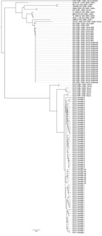
Figure 3. Core genome phylogeny illustrating the evolutionary history of the of Shiga toxin–producing Escherichia coli (STEC) O55 strain from the July 2014 Dorset County, England, outbreak in the context of STEC O157:H7...
To investigate the evolutionary history of the STEC O55 Dorset outbreak strain, we constructed a core genome phylogeny (Figure 3). The analysis divided the sequences of the isolates in this study according to serotype; all isolates of E. coli O55:H7 clustered together on a separate branch of the tree, and all isolates of STEC O157:H7 clustered together on the branch below, regardless of sorbitol/β-glucuronidase phenotype or the presence of stx (Figure 3). The phylogenetic analysis of E. coli O55:H7 indicated that incorporation of the Stx-encoding prophage has occurred on multiple occasions within the EPEC O55:H7 background, with independent acquisition of stx1 (15), stx2d, and stx2a into EPEC O55:H7. Likewise, multiple acquisition and loss events involving stx1, stx2c, stx2a, and less commonly stx2d have been described for STEC O157:H7 (12,32).
As noted by McFarland et al. (1), the outbreak strain was closely related to STEC O55:H7 stx2a isolates identified in Ireland during 2013–2014 (Figure 3). These 6 isolates from Ireland were <5 SNPs from the Dorset outbreak strain, indicating that the isolates from Ireland and Dorset County shared a common source (8). The outbreak strain had lost the ability to ferment sorbitol, which appears to be a recent adaption with all ancestral O55:H7, including those isolated in Ireland in 2012 retaining the ability to ferment sorbitol. A similar relationship exists between the sorbitol-positive and sorbitol-negative STEC O157:H7 phenotypes; the sorbitol-negative phenotype is a more recent adaption from the sorbitol-positive progenitor strain (Figure 3) (3,5).
The most parsimonious model of evolution of the STEC O55:H7 Dorset outbreak strain was a progenitor EPEC O55:H7 sorbitol-fermenting strain lysogenized by an Stx2a-encoding phage and subsequent loss of the ability to ferment sorbitol. This stepwise model of evolution seems to mirror that seen in the common STEC O157:H7 clone; the acquisition of the STEC pathotype preceded phenotypic modulation.
In the United Kingdom, STEC is regarded as a substantial threat to public health, and enhanced surveillance systems are in place (32). In England, HUS developed in ≈5% of symptomatic STEC O157:H7 patients (33), notably less than the 42% of patients in whom HUS developed during the STEC O55:H7 outbreak described in this study. The Dorset outbreak strain was closely related to the common STEC O157:H7 clone and shared several characteristics, most notably the presence of phage-encoded stx2a. Stx2a is associated with more severe symptoms, including the development of HUS, and it is probably the key virulence factor causing the high proportion of HUS cases in this outbreak (10). Of additional concern was the inability to detect the outbreak strain at the local hospital level by using the standard microbiology investigation method, cefixime and tellurite sorbitol MacConkey agar, because of this strain’s sensitivity to tellurite.
A previously published stepwise evolutionary model showed the acquisition of stx2 by a strain of EPEC O55:H7, resulting in emergence of a strain of STEC O55:H7, which was β-glucuronidase positive and sorbitol positive, closely related but ancestral to STEC O157:H7, which was β-glucuronidase positive and sorbitol positive (34). The loss of the sorbitol-positive phenotype in STEC O157:H7 was followed by the loss of β-glucuronidase expression, resulting in the common STEC O157 sorbitol-negative β-glucuronidase–negative clone. The evolutionary history of the Dorset outbreak strain begins with the EPEC O55:H7 progenitor strain described previously (6) (Figure 3). Subsequent acquisition of an Stx2a-encoding phage was confirmed by detection of STEC O55:H7 β-glucuronidase–positive sorbitol-positive isolates in Ireland in 2012 (Figure 3). The loss of the sorbitol-positive phenotype mirrored the genetic events proposed to have occurred in the evolution of STEC O157, albeit by an alternative mechanism.
The parallel, convergent evolutionary history of STEC O157:H7 and STEC O55:H7 may indicate a common driver in the evolutionary process. Adaptation to a new niche may be accompanied by modification of gene expression because genes no longer required for, or incompatible with, the variation in lifestyle are selectively inactivated by point mutation, insertion, or deletion (35). Loss of the sorbitol-positive phenotype may coincide with the successful colonization of a new animal host or the ability to transmit more effectively between animal hosts without the need to survive in the environment for long periods (28,36).
The detection of the STEC O55:H7 sorbitol-negative strain in patients in Ireland before the outbreak in Dorset led to speculation that ruminants (most likely cattle or sheep) on the island of Ireland were the source of the outbreak strain (1). Transmission between Ireland and Dorset may have occurred via movement of persons, livestock, or a secondary vector such as migratory birds (37). The finding that the Stx2a-encoding phage has a high level of similarity to Stx2a-encoding phage found in a previously described sublineage of STEC O157 PT32 geographically linked to Ireland may provide further evidence of the origin of this strain (17,29). Phages from STEC O157 may be exchanged with other phages from serotypes of E. coli in the gut of the ruminant host or in the environment. Analysis and comparison of phage sequences to provide clues regarding the origin of a strain of STEC is a novel approach to outbreak investigation; additional studies are required to evaluate the utility of the approach. Further work will be hampered by the lack of available sequences of the Stx-encoding phage and the difficulties with assembling the sequences because of the inability of short-read sequencing to resolve the large number of repetitive and paralogous features characteristic of the prophage.
The STEC O55:H7 Dorset outbreak strain described in this study shared characteristics with the common STEC O157:H7 clone, specifically the acquisition of an Stx2a-encoding phage and the sorbitol-negative phenotype. Key differences between the 2 strains include the rfb gene cluster, plasmid content, β-glucuronidase phenotype, and the absence of the ter gene cluster in the STEC O55:H7 outbreak strain. Despite these differences, this study provides evidence of parallel, convergent evolution of STEC O157:H7 and STEC O55:H7, involving multiple acquisitions of Stx-encoding phages and loss of the ability to ferment sorbitol. Previous studies have shown a clear association with STEC harboring stx2a and progression to HUS (10). Acquisition of the Stx2a-encoding phage seems to explain the emergence of STEC O157:H7 as a clinically significant pathogen; in contrast to the acquisition of stx2c, evidence suggests that after Stx2a-encoding phage is integrated in a population, it tends to be maintained and may be associated with higher excretions levels in cattle (29,36). As such, the emergence of STEC O55:H7 harboring stx2a is of public health concern.
Mr. Schutz is a recent graduate of the London School of Hygiene and Tropical Medicine and the Royal Veterinary College in London. His research interests include emerging zoonoses and host adaptation.
Acknowledgments
We acknowledge all members of the Outbreak Control Team, especially Noeleen McFarland, Amy Mikhail, and Sooria Balasegaram.
This work was supported by the National Institute for Health Research Health Protection Research Unit in Gastrointestinal Infections. D.L.G., T.J.D., and S.S. were supported by a Food Standards Scotland/Food Standards Agency study grant (FS101055).
References
- McFarland N, Bundle N, Jenkins C, Godbole G, Mikhail A, Dallman T, et al. Recurrent seasonal outbreak of an emerging serotype of Shiga toxin-producing Escherichia coli (STEC O55:H7 Stx2a) in the south west of England, July 2014 to September 2015. Euro Surveill. 2017;22:30610. DOIPubMedGoogle Scholar
- Whittam TS, Wolfe ML, Wachsmuth IK, Orskov F, Orskov I, Wilson RA. Clonal relationships among Escherichia coli strains that cause hemorrhagic colitis and infantile diarrhea. Infect Immun. 1993;61:1619–29.PubMedGoogle Scholar
- Feng P, Lampel KA, Karch H, Whittam TS. Genotypic and phenotypic changes in the emergence of Escherichia coli O157:H7. J Infect Dis. 1998;177:1750–3. DOIPubMedGoogle Scholar
- Iguchi A, Ooka T, Ogura Y, Asadulghani , Nakayama K, Frankel G, et al. Genomic comparison of the O-antigen biosynthesis gene clusters of Escherichia coli O55 strains belonging to three distinct lineages. Microbiology. 2008;154:559–70. DOIPubMedGoogle Scholar
- Wick LM, Qi W, Lacher DW, Whittam TS. Evolution of genomic content in the stepwise emergence of Escherichia coli O157:H7. J Bacteriol. 2005;187:1783–91. DOIPubMedGoogle Scholar
- Zhou Z, Li X, Liu B, Beutin L, Xu J, Ren Y, et al. Derivation of Escherichia coli O157:H7 from its O55:H7 precursor. PLoS One. 2010;5:e8700. DOIPubMedGoogle Scholar
- Kyle JL, Cummings CA, Parker CT, Quiñones B, Vatta P, Newton E, et al. Escherichia coli serotype O55:H7 diversity supports parallel acquisition of bacteriophage at Shiga toxin phage insertion sites during evolution of the O157:H7 lineage. J Bacteriol. 2012;194:1885–96. DOIPubMedGoogle Scholar
- Dallman TJ, Byrne L, Ashton PM, Cowley LA, Perry NT, Adak G, et al. Whole-genome sequencing for national surveillance of Shiga toxin-producing Escherichia coli O157. Clin Infect Dis. 2015;61:305–12. DOIPubMedGoogle Scholar
- Jenkins C, Lawson AJ, Cheasty T, Willshaw GA. Assessment of a real-time PCR for the detection and characterization of verocytotoxigenic Escherichia coli. J Med Microbiol. 2012;61:1082–5. DOIPubMedGoogle Scholar
- Persson S, Olsen KE, Ethelberg S, Scheutz F. Subtyping method for Escherichia coli shiga toxin (verocytotoxin) 2 variants and correlations to clinical manifestations. J Clin Microbiol. 2007;45:2020–4. DOIPubMedGoogle Scholar
- Stevens MP, Frankel GM. The locus of enterocyte effacement and associated virulence factors of enterohemorrhagic Escherichia coli. Microbiol Spectr. 2014;2:EHEC-0007–2013. http://dx.doi.org: DOIGoogle Scholar
- Feng P, Lampel KA. Genetic analysis of uidA expression in enterohaemorrhagic Escherichia coli serotype O157:H7. Microbiology. 1994;140:2101–7. DOIPubMedGoogle Scholar
- Lim JY, Yoon J, Hovde CJ. A brief overview of Escherichia coli O157:H7 and its plasmid O157. J Microbiol Biotechnol. 2010;20:5–14.PubMedGoogle Scholar
- Mainda G, Lupolova N, Sikakwa L, Bessell PR, Muma JB, Hoyle DV, et al. Phylogenomic approaches to determine the zoonotic potential of Shiga toxin-producing Escherichia coli (STEC) isolated from Zambian dairy cattle. Sci Rep. 2016;6:26589. DOIPubMedGoogle Scholar
- Lindsey RL, Rowe L, Garcia-Toledo L, Loparev V, Knipe K, Stripling D, et al. High-quality draft genome sequences for five non-o157 Shiga toxin-producing Escherichia coli strains generated with PacBio sequencing and optical maps. Genome Announc. 2016;4:e00626–16. DOIPubMedGoogle Scholar
- Hayashi T, Makino K, Ohnishi M, Kurokawa K, Ishii K, Yokoyama K, et al. Complete genome sequence of enterohemorrhagic Escherichia coli O157:H7 and genomic comparison with a laboratory strain K-12. DNA Res. 2001;8:11–22. DOIPubMedGoogle Scholar
- Shaaban S, Cowley LA, McAteer SP, Jenkins C, Dallman TJ, Bono JL, et al. Evolution of a zoonotic pathogen: investigating prophage diversity in enterohaemorrhagic Escherichia coli O157 by long-read sequencing. Microb Genom. 2016;2:e000096.PubMedGoogle Scholar
- Bankevich A, Nurk S, Antipov D, Gurevich AA, Dvorkin M, Kulikov AS, et al. SPAdes: a new genome assembly algorithm and its applications to single-cell sequencing. J Comput Biol. 2012;19:455–77. DOIPubMedGoogle Scholar
- Seemann T. Prokka: rapid prokaryotic genome annotation. Bioinformatics. 2014;30:2068–9. DOIPubMedGoogle Scholar
- Darling AE, Mau B, Perna NT. progressiveMauve: multiple genome alignment with gene gain, loss and rearrangement. PLoS One. 2010;5:e11147. DOIPubMedGoogle Scholar
- Altschul SF, Gish W, Miller W, Myers EW, Lipman DJ. Basic local alignment search tool. J Mol Biol. 1990;215:403–10. DOIPubMedGoogle Scholar
- Zhou Y, Liang Y, Lynch KH, Dennis JJ, Wishart DS. PHAST: a fast phage search tool. Nucleic Acids Res. 2011;39(suppl):W347-52. DOIPubMedGoogle Scholar
- Alikhan NF, Petty NK, Ben Zakour NL, Beatson SA. BLAST Ring Image Generator (BRIG): simple prokaryote genome comparisons. BMC Genomics. 2011;12:402. DOIPubMedGoogle Scholar
- Bolger AM, Lohse M, Usadel B. Trimmomatic: a flexible trimmer for Illumina sequence data. Bioinformatics. 2014;30:2114–20. DOIPubMedGoogle Scholar
- Li H, Durbin R. Fast and accurate long-read alignment with Burrows-Wheeler transform. Bioinformatics. 2010;26:589–95. DOIPubMedGoogle Scholar
- McKenna A, Hanna M, Banks E, Sivachenko A, Cibulskis K, Kernytsky A, et al. The Genome Analysis Toolkit: a MapReduce framework for analyzing next-generation DNA sequencing data. Genome Res. 2010;20:1297–303. DOIPubMedGoogle Scholar
- Stamatakis A. RAxML version 8: a tool for phylogenetic analysis and post-analysis of large phylogenies. Bioinformatics. 2014;30:1312–3. DOIPubMedGoogle Scholar
- Ingle DJ, Tauschek M, Edwards DJ, Hocking DM, Pickard DJ, Azzopardi KI, et al. Evolution of atypical enteropathogenic E. coli by repeated acquisition of LEE pathogenicity island variants. Nat Microbiol. 2016;1:15010. DOIPubMedGoogle Scholar
- Dallman TJ, Ashton PM, Byrne L, Perry NT, Petrovska L, Ellis R, et al. Applying phylogenomics to understand the emergence of Shiga-toxin-producing Escherichia coli O157:H7 strains causing severe human disease in the UK. Microb Genom. 2015;1:e000029. DOIPubMedGoogle Scholar
- Feng PC, Jinneman K, Scheutz F, Monday SR. Specificity of PCR and serological assays in the detection of Escherichia coli Shiga toxin subtypes. Appl Environ Microbiol. 2011;77:6699–702. DOIPubMedGoogle Scholar
- Monday SR, Whittam TS, Feng PC. Genetic and evolutionary analysis of mutations in the gusA gene that cause the absence of beta-glucuronidase activity in Escherichia coli O157:H7. J Infect Dis. 2001;184:918–21. DOIPubMedGoogle Scholar
- Sánchez S, Llorente MT, Herrera-León L, Ramiro R, Nebreda S, Remacha MA, et al. Mucus-activatable Shiga toxin genotype stx2d in Escherichia coli O157:H7. Emerg Infect Dis. 2017;23:1431–3. DOIPubMedGoogle Scholar
- Byrne L, Jenkins C, Launders N, Elson R, Adak GK. The epidemiology, microbiology and clinical impact of Shiga toxin-producing Escherichia coli in England, 2009-2012. Epidemiol Infect. 2015;143:3475–87. DOIPubMedGoogle Scholar
- Feng PC, Monday SR, Lacher DW, Allison L, Siitonen A, Keys C, et al. Genetic diversity among clonal lineages within Escherichia coli O157:H7 stepwise evolutionary model. Emerg Infect Dis. 2007;13:1701–6. DOIPubMedGoogle Scholar
- Maurelli AT. Black holes, antivirulence genes, and gene inactivation in the evolution of bacterial pathogens. FEMS Microbiol Lett. 2007;267:1–8. DOIPubMedGoogle Scholar
- Matthews L, Reeve R, Gally DL, Low JC, Woolhouse ME, McAteer SP, et al. Predicting the public health benefit of vaccinating cattle against Escherichia coli O157. Proc Natl Acad Sci U S A. 2013;110:16265–70. DOIPubMedGoogle Scholar
- See comment in PubMed Commons belowPersad AK, LeJeune JT. Animal reservoirs of Shiga toxin-producing Escherichia coli. Microbiol Spectr. 2014;2:EHEC-0027–2014.
Figures
Tables
Cite This ArticleTable of Contents – Volume 23, Number 12—December 2017
| EID Search Options |
|---|
|
|
|
|
|
|
Please use the form below to submit correspondence to the authors or contact them at the following address:
Claire Jenkins, Gastrointestinal Bacteria Reference Unit, Public Health England, 61 Colindale Ave, NW9 5HT, UK
Top