Volume 23, Number 6—June 2017
Dispatch
Central Nervous System Brucellosis Granuloma and White Matter Disease in Immunocompromised Patient
Mohammed Alqwaifly , Fahad S. Al-Ajlan, Hindi Al-Hindi, and Abdulaziz Al Semari
Figure 2
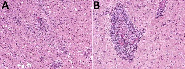
Figure 2. Histologic analysis of a brain biopsy specimen from a 46-year-old immunocompromised woman with central nervous system brucellosis granuloma and white matter disease, Saudi Arabia. A) Low magnification view of cerebral cortex showing infiltration by perivascular lymphocytes and histiocytes. Histiocytes form small nonnecrotizing granuloma (center) (original magnification ×100). B) High magnification view showing an angiocentric epithelioid granuloma cuffed by mature lymphocytes (original magnification ×200). Hemotoxylin and eosin stain.
Page created: May 16, 2017
Page updated: May 16, 2017
Page reviewed: May 16, 2017
The conclusions, findings, and opinions expressed by authors contributing to this journal do not necessarily reflect the official position of the U.S. Department of Health and Human Services, the Public Health Service, the Centers for Disease Control and Prevention, or the authors' affiliated institutions. Use of trade names is for identification only and does not imply endorsement by any of the groups named above.