Volume 24, Number 1—January 2018
Research Letter
Emmonsia helica Infection in HIV-Infected Man, California, USA
Martin Rofael , Ilan S. Schwartz, Lynne Sigler, Li K. Kong, and Nicholas Nelson
Figure
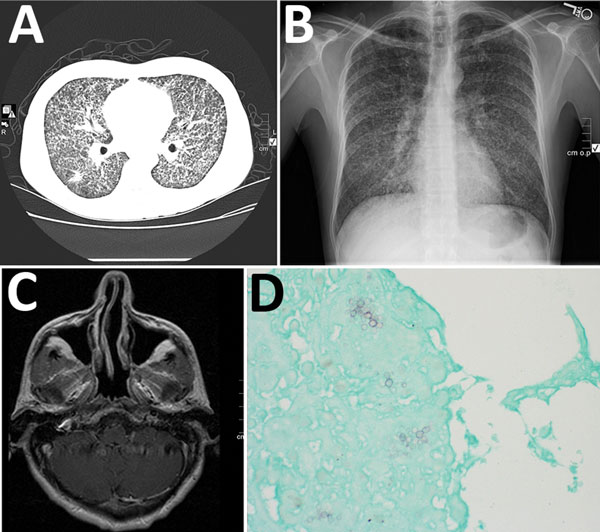
Figure. Emmonsia helica infection in an immunocompromised man, California, USA, 2016. A) Chest radiograph with diffuse micronodularities throughout both lung fields. B) Computed tomographic scan with diffuse micronodular pulmonary disease. C) Axial magnetic resonance image with 6-mm ring-enhancing lesion in the right cerebellum adjacent to the fourth ventricle. D) Grocott’s methenamine-silver stain showing broad-based budding yeast. Original magnification ×400.
Page created: January 24, 2018
Page updated: January 24, 2018
Page reviewed: January 24, 2018
The conclusions, findings, and opinions expressed by authors contributing to this journal do not necessarily reflect the official position of the U.S. Department of Health and Human Services, the Public Health Service, the Centers for Disease Control and Prevention, or the authors' affiliated institutions. Use of trade names is for identification only and does not imply endorsement by any of the groups named above.