Volume 24, Number 12—December 2018
Research Letter
Disseminated Spiroplasma apis Infection in Patient with Agammaglobulinemia, France
Nicolas Etienne, Laurent Bret, Cécile Le Brun, Hervé Lecuyer, Josquin Moraly, Fanny Lanternier, Olivier Hermine, Agnès Ferroni, Marc Lecuit, Sabine Pereyre1, Laure Beven1, and Olivier Lortholary1
Figure
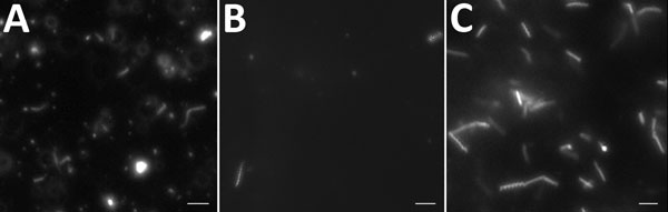
Figure. Direct examination with dark-field microscopy of specimens from a patient with agammaglobulinemia who had Spiroplasma apis infection, France. A) Helical and motile bacteria in blood culture. B) Elongated and coccoid bacteria in joint fluid. C) Helical and motile bacteria in culture from joint fluid in modified SP4 broth medium. Scale bar indicates 10 µm.
1These authors contributed equally to this article.
Page created: November 20, 2018
Page updated: November 20, 2018
Page reviewed: November 20, 2018
The conclusions, findings, and opinions expressed by authors contributing to this journal do not necessarily reflect the official position of the U.S. Department of Health and Human Services, the Public Health Service, the Centers for Disease Control and Prevention, or the authors' affiliated institutions. Use of trade names is for identification only and does not imply endorsement by any of the groups named above.