Volume 24, Number 2—February 2018
Research Letter
Amebaborne “Attilina massiliensis” Keratitis, France
Figure

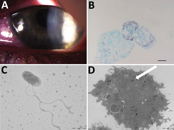
Figure. Results of testing for a 17-year-old woman with keratoconjunctivitis symptoms, Marseille, France, July 2016. A) Slit-lamp optic microscopic photograph of left eye infected with pseudo-dendritic keratitis associated with Acanthamoeba castellani–“Attilina massiliensis” ocular infection. B) Microscopic aspect of A. castellani ameba infected by “A. massiliensis” from corneal swab sample. Scale bar indicates 1 μm. C) Optic microscopy image of flagellated, free-living “A. massiliensis” from swab sample. Scale bar indicates 1 μm. D) Electron microscopy image of the ameba containing the “A. massiliensis” endosymbiont, stained by using Gimenez staining (white arrow). Scale bar indicates 2 μm.
Page created: January 17, 2018
Page updated: January 17, 2018
Page reviewed: January 17, 2018
The conclusions, findings, and opinions expressed by authors contributing to this journal do not necessarily reflect the official position of the U.S. Department of Health and Human Services, the Public Health Service, the Centers for Disease Control and Prevention, or the authors' affiliated institutions. Use of trade names is for identification only and does not imply endorsement by any of the groups named above.