Volume 24, Number 2—February 2018
Research Letter
Cerebral Syphilitic Gumma in Immunocompetent Man, Japan
Tatsuya Kodama , Hidenori Sato, Morichika Osa, Yuji Fujikura, and Akihiko Kawana
Figure
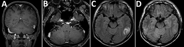
Figure. Brain magnetic resonance imaging findings in a 36-year-old immunocompetent man before (A, B, C) and after (D) treatment for cerebral syphilitic gumma, Saitama, Japan. A) Gadolinium-enhanced T1-weighted coronal image shows an enhanced nodular lesion in the left temporal lobe. B) Axial gadolinium-enhanced T1-weighted image shows enhancement within the cisternal segment of both the vestibulocochlear nerve complex and the facial nerve. C) Axial fluid-attenuated inversion recovery images show a hyperintense lesion-like mass in the left temporal lobe. D) Axial fluid-attenuated inversion recovery image shows complete resolution after discontinuation of treatment.
Page created: January 17, 2018
Page updated: January 17, 2018
Page reviewed: January 17, 2018
The conclusions, findings, and opinions expressed by authors contributing to this journal do not necessarily reflect the official position of the U.S. Department of Health and Human Services, the Public Health Service, the Centers for Disease Control and Prevention, or the authors' affiliated institutions. Use of trade names is for identification only and does not imply endorsement by any of the groups named above.