Volume 24, Number 2—February 2018
Research Letter
Human African Trypanosomiasis in Emigrant Returning to China from Gabon, 2017
Xinyu Wang1, Qiaoling Ruan1, Wen-Hong Zhang1, Jianfei Gu, Yiyi Qian, Muxin Chen, Qin Liu, Qing Lu, and Wenhong Zhang
Figure
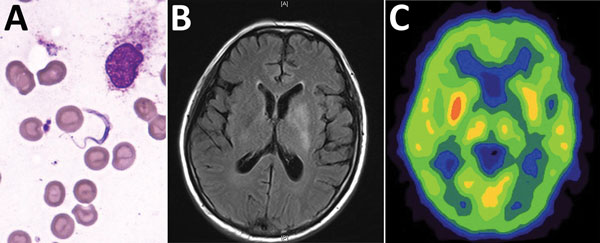
Figure. Bone marrow test results and brain imaging of a 60-year-old man who returned to China from Gabon with suspected human African trypanosomiasis. A) Trypanosoma spp. (later determined to be T. brucei gambiense) in a Giemsa-stained thin bone marrow film. Original magnification ×1,000. B) A T2-weighted fluid-attenuated inversion recovery image with hyperintense signal changes in the left basal ganglia. C) Brain positron emission tomography–computed tomography suggested reduced glucose metabolism in the left basal ganglia.
1These authors contributed equally to this article.
Page created: January 17, 2018
Page updated: January 17, 2018
Page reviewed: January 17, 2018
The conclusions, findings, and opinions expressed by authors contributing to this journal do not necessarily reflect the official position of the U.S. Department of Health and Human Services, the Public Health Service, the Centers for Disease Control and Prevention, or the authors' affiliated institutions. Use of trade names is for identification only and does not imply endorsement by any of the groups named above.